Wednesday, September 27, 2023

When Immunity Fails Programmed Cell Death

DNA Damage Response

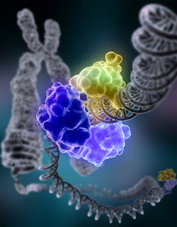
Telomeric repeat (TR) sequences are responsible for genome integrity, where instability is a primary factor that leads to activation of p53. Introduction of a TR into cells leads to stabilization of p53, specific to TRs and not observed in plasmids containing non-TR sequences. TR-activated p53 exhibited enhanced transcriptional activity and induced p53-dependent growth suppression, measured as a reduction in colony formation. Sub-telomeric p53 binding prevents accumulation of DNA damage at human telomeres.  

Healthy cells experience thousands of DNA lesions per day. Micronuclei, containing broken fragments of DNA or chromosomes, that have become isolated, are recognized as one mediator of DNA damage response (DDR)-associated immune recognition. Like micronuclear DNA, mitochondrial DNA (mtDNA) is recognized by cGAS to drive STING-mediated inflammatory signaling. Mitochondrial damage can intersect DNA repair and inflammatory cascades with programmed cell death, through p53. In human fibroblasts and conditionally immortalized vascular smooth muscle cells p53 mediates CD54 (ICAM-1) overexpression in senescence.

Replicative senescence, an autophagy dependent program and crisis are anti-proliferative barriers that human cells must evade to gain immortality. Telomere-to-mitochondria signaling by ZBP1 mediates replicative crisis. Dysfunctional telomeres activate innate immune responses (IFN) through mitochondrial TR RNA (TERRA)–ZBP1 complexes. Senescence occurs when shortened telomeres elicit a p53 and RB dependent DNA-damage response. A crisis-associated isoform of ZBP1(innate immune sensor) is induced by the cGAS–STING DNA-sensing pathway, but reaches full activation only when associated with TERRA transcripts from dysfunctional telomeres. p53 utilizes the cGAS/STING innate immune system pathway for both cell intrinsic and cell extrinsic tumor suppressor activities. cGAS-STING activation induces the production of IFN-b and increases CD54 expression in  human cerebral microvascular endothelial cells.

In melanoma patients there is a significant correlation between cGAS expression levels and survival and between NK cell receptor expression levels and survival. Loss of cGAS expression by tumor cells could permit the tumor cell to circumvent senescence or prevent immunostimulatory NKG2D ligands expression. Loss of p53 and gain of oncogenic RAS exacerbated pro-malignant paracrine signaling activities of senescence-associated secretory phenotypes. Results imply that heterogeneity in cGAS activity, across tumors, could be an important predictor of cancer prognosis and response to treatment and suggest that NK cells could play an important role in mediating anti-tumor effects. Coculture of wild-type p53-induced human tumor cells with primary human NK cells enhanced NKG2D-dependent degranulation and IFN-γ production by NK cells. 

When p53 consensus sequences are modified and DNA damage response is compromised, replicative crisis ensues, mitochondrial membranes misfunction, mtDNA expression is downregulated and IFN signaling upregulates. A cell may then express activating immune ligands that bind NK receptors signaling non-self and cytolytic death or inhibitory receptors that signal self and immortality


Thursday, September 21, 2023

Indispensable Mitochondria - Cancers back door?


Immediately prior to fertilization spermatozoa are devoid of Mitochondrial DNA (mtDNA), potentially explaining an aspect about selection that may serve the legacy for maternal immune tolerance. Post fertilization, on day 11-13, outermost trophoblasts of the blastocyst dock with the decidual lining as it embeds in the uterine wall. Then, maternal vascular remodeling and placental formation begin toward successful implantation. 

Higher quality trophoblasts are associated with lower mtDNA content. Moreover, euploid blastocysts with higher mtDNA content had a lower chance to implant and mtDNA replication is strictly downregulated between fertilization and the implantation. What is it about absent or reduced mtDNA that may also relate to the mechanics of immune tolerance and vascular remodeling, which are also features of solid tumors.

The initial absence or downregulation of MtDNA, may relate an immune tolerance by uterine Natural Killer (NK) cells. As mtDNA upregulates, after day 12, it may initiate NK auto-reactivity required for maternal microvascular remodeling. This auto-immune paradox is a prerequisite for vascular remodeling, which is also seen in localized hypertension, and the likely basis of successful blastocyst implantation. Acutely, micro-hypertension induced mechanical stretch, on endothelial cells, interconnects innate and adaptive immune responses. 

The dominant cell in the decidua is an NK subset (dNK), they express low levels of IFN-γ and express proteins of Renin Angiotensin System (RAS). At day 12 RAS peptide ANP colocalizes to dNK’s suggesting that dNK RAS infers localized responsiveness.  When TFAM, required for transcription of mtDNA, was deleted from cardiomyocytes, after 32 days, animals developed cardiomyopathy and Nppa (gene encoding ANP) and Nppb expression was elevated. 

In monocytes increased endothelial stretch activates STAT3, which is involved in driving almost all pathways that control NK cytolytic activity and reciprocal regulatory interactions between NK cells and other components of the immune system. The crosstalk between STAT3 and p53/RAS signaling controls cancer cell metastasis. p53, Stat3, and, potentially, the estrogen receptor are thought to act as co-regulators, affecting mitochondrial gene expression through protein-protein interactions. Co-immunoprecipitation of p53 with TFAM suggests it may regulate mitochondrial DNA-damage repair.

Like initial trophoblasts with low level mtDNA, mature cells, like cardiomyocytes that prolong low level mtDNA may also aggravate autoimmune sponsored hypertension that remodels microvascular networks providing nutrients for growth of reduced mtDNA stem cell replicas. Indeed, mitochondrial dysfunction (from depleted mtDNA) does not affect pluripotent gene expression, but results in severe defects in lineage differentiation.

During severe sepsis, intense, on-going mtDNA damage and mitochondrial dysfunction could overwhelm the capacity for mitochondrial biogenesis, leading to a gradual decline in mtDNA levels over time. This may contribute to monocyte immune deactivation, which is associated with adverse clinical outcomes and could be reversed by IFN-γ

Identifying cells that optimally educate cocultured NK cells for precision IFN-γ and cytolytic responsiveness is part of the ongoing work by the Codondex team.