Monday, December 28, 2020

Natural Killers at the Neuro-Immune Axis!

Much has been said about the role Natural Killer (NK) cells play in positively and negatively influencing events in tissues and cells. Summarized facts about the healthy state of NK cells in humans and animals explain how innate immune cells, including NK cells differ from adaptive immune cells. One significant feature of NK cells is that they can act independently of MHC antigen presentations and that makes them tantalizing, but enormously challenging for scientists seeking to embrace their  influence over cells and their killing capabilities.

The variety and combination of inhibitory and activating receptors differentially expressed by as many as 30,000 human NK cell subsets makes heterogeneity difficult to relate across different conditions, organs and tissue types. Notwithstanding, positive rates of overall patient survival resoundingly corelated to the presence of as few as one NK cell infiltrating a tumor in a microscopic field.  

Innate immune cells including NK and macrophages have also been directly tied to conditions of neurological pain and more specifically to afferent and efferent fibers that signal through the vagus nerve. In these models at the immune-neurological interface similarities exists and both organs must interact for proper function. 

In each of these organs communication is mediated by direct cellular contact eg. synapse formation and via soluble mediators like cytokines or neurotransmitters that also communicate bi-directionally between cells of each system. The nervous and immune systems can influence each other’s activity because immune cells express neurotransmitter receptors, and neurons express cytokine receptors. Immune cells can synthesize and release neurotransmitters themselves, thus using neurotransmitter-mediated pathways via autocrine and paracrine mechanisms. This may indicate that NK cells extend nerve end signaling further into tissues and at a cellular level. 

A recent paradigm in physiology describes the existence of neuro-immune cell units, at an organ-tissue level and identifies the enormous complexity inherent in this globally unifying approach that also connects neuro-immune-gut, at least in Parkinson's disease.

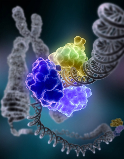
Parkinson’s is a brain disorder where certain nerve cells slowly die and symptoms worsen. The risk of developing the condition increases with age, but in certain patients the illness is caused by defects in two proteins, PINK1 and parkin. NK cells are capable of homing to the central nervous system in neurological disorders that exhibit exacerbated inflammation and inhibit hyperactivated microglia. Recently, a study demonstrated that NK cells scavenge alpha-synuclein aggregates and systemic depletion of NK cells results in exacerbated neuropathology in a mouse model of alpha-synucleinopathy, making NK cells highly relevant in Parkinson’s disease.

We recently described a mechanism by which the sentinel state of NK cells is impaired and suggested the senescent phenotype, induced by age related mitophagy could be the primary cause. Increase in mitophagy (mitochondrial autophagy) is age-dependent and abrogated by PINK1 or parkin deficiency suggesting, in Parkinson's disease compromised mitophagy is associated with neurological degeneration. Further  PINK1 and Parkin, which are regulated by p53 specifically repress mitochondrial antigen presentation of both MHC classes. Therefore, excessive PINK1 or parkin increases rates of NK cell mitophagy and repress the presentation of mitochondrial antigens for MHC classes at the axis of this neuro-immune related disease.

The healthy state of NK cells at the axis of neuro-immune systems may indeed have more far reaching implications for the future of human diseases and therapies.


Sunday, December 13, 2020

Natural Killers Linked to Overall Survival in Cancer

A meta analysis of tumor samples, collected between 1973 and 2016, in 53 studies confirmed overall survival (OS) correlated with Natural Killer cell infiltration into solid tumors. The number of NK cells infiltrating solid tumors, including those considered “highly ”infiltrated was relatively low, compared with other immune populations. Notwithstanding, the presence of a single NK cell, within a high powered microscopic field was associated with significantly improved OS and disease free survival in colorectal cancer, HER2 + breast cancer and hepatocellular carcinoma.

The finding supports the prospect that single tumor infiltrating NK cells, in a sampled tissue can be determinative for OS. By inference a single tumor infiltrating NK cell or cells possess characteristics that are relative to OS and beneficial to patient.  

NK cell surface receptors are densely varied defining at least 30,000 unique NK cell populations within each individual. NK cell classifications, relative to tumor infiltration and OS is enormously complex, especially at this scale and present definitions of activating and inhibiting receptor combinations underwhelm. To identify NK cells that have infiltrated or may be capable of infiltrating a patient tumor to improve OS we focused on biopsied tumor tissue selections whether or not they include NK cells.

Our work is with two tumor types in humanized mice. Multiple sections of each tumor were resected and divided into multiple parts for coculture with allogenic naïve, IL2 and probiotic enhanced NK cells and for DNA sequencing. After coculture NK cell cytotoxicity and other detailed measures resulting from each resected section and from single cells were assessed. Presently sequencing of DNA from each resected, divided section (pre-coculture) is focused on comparisons derived from TP53.

In the final stage NK cells will be cocultured with resected tumor tissue and will be made to challenge new tumor tissue and single cells, from the resected tumor from which the NK coculture was derived. The objective will be whether Codondex analysis of TP53 DNA sequencing can predict the most successful tumor tissue candidates based upon the most effective cocultured NK cell challenge to the tumor derived tissue or cells. 

If Codondex algorithm is found to identify a direct or indirect logic for tissue or cell selection that is effective in vitro our work will continue to next stage in vivo testing and analysis on similar grounds. 


Wednesday, November 25, 2020

Not Only A Killer A System for Killing!

The next time you're out exercising, spare a thought for your busy mitochondria. NASA scientists just reported mitochondria as the key to health problems in space.

Natural killer (NK) cells can extend membrane probes into cells or pathogens. These are loaded with granulysin (GNLY) to penetrate and perforin (PFN) to kill intracellular bacteria or protozoa and can lyse entire cells. The probes can also transfer healthy mitochondria to apoptotic cardiomyocytes (and other cells) in need of mitochondrial transfer. Uterine NK cells of the decidua send probes into trophoblasts to selectively kill intracellular Listeria monocytogenes without killing the trophoblast host. Stressed cells, moving toward apoptosis can behave similarly, but in reverse shooting out nanoprobes to proximal cells seeking cooperation and urgent mitochondrial transfers including to cancer cells.

A meta-analysis of gene expression signatures for blood pressure and hypertension in 7017 individuals from 6 international studies found of 7717 genes, 34 were most differentialy expressed including GNLY. Enrichment analysis for the diastolic and systolic gene group's associated strongly with NK cell mediated cytotoxicity and 13 other pathways including antigen processing and inflammatory response.

Formation of membrane probes or tubes, in which mitochondria travel and establishment of intracellular mitochondrial networks in the peripheral zone of cells require Kinesin-1 heavy chain (KIF5B). KIF5B is also required for female meiosis (oogenesis) and proper chromosomal segregation in mitotic cells and modulates central spindle organization in late-stage cytokinesis in chondrocytes.

A study of centromere heterochromatin (connected with central spindle) surprisingly showed that distant euchromatic regions, enriched in repressed methylated genes also interacted with the hierarchical organization of centromeric DNA. These 3D spatial interactions (at a distance) are likely mediated by liquid-like fusion events and can influence the health of individuals. Repressed gene's were identified as transposable elements, sequences often associated with pathogenic DNA insertions that have been persistently retained.  

KIF5B is an interaction partner of ADP-ribosylation factor-like 8b (Arl8b), which is required for NK cell–mediated cytotoxicity that drives polarization of lytic granules and microtubule-organizing centers (MTOCs) toward the immune synapse between NK and target cells. Silencing experiments that led to failure of MTOC-lytic granule polarization suggest Arl8b and KIF5B together control the critical step in NK cell cytotoxicity. 

KIF5B is also a critical transporter of p53 and c-Myc to the cytoplasm for degradation. However, subcellular localization of Arl8b and p53-dependent cell death was shown to occur through knockdown of acetylation subunit NatC. As a consequence, p53 is stabilized, phosphorylated and significantly activates transcription of downstream proapoptotic genes. In the absence KIF5B, or presence of  mutants p53 and c-Myc aggregate in the nucleus where they signal DNA damage-induced apoptosis through the control of p53 by endogenous c-Myc (in vivo).

Finely tuned, frequently used KIF5B in NK cells expressing GNLY may induce effects on local tissue blood pressure, as was discovered by expression of Renin-Angiotensin vasoactive proteins AT1, AT2, and ANP in pregnancy-induced uterine NK cellsInflammation signaling, via tissue bound NK cells may result from stretch-mediated release of angiotensin II, which is coupled with p53 acetylation apoptosis and activation of p53. This may prolong upregulation of the local renin-angiotensin system, increase susceptibility of target cells to apoptosis and signal adaptive immune cells. 

Somewhere in the balance between NatC knockdown induced apoptosis and angiotensin II induced apoptosis p53 may direct traffic to keep your cells healthy!


Monday, November 2, 2020

An Integrated P53 Puzzle - Glycolysis in Cancer, Diabetes and Immunity!

Oxygen poor, hypoxic tissue promotes a cellular shift in mitochondrial metabolism from OXPHOS to less energy efficient glycolysis. Each shift induces environmental, epigenetic and genetic factors that alter a cells response to insult, attack and disease. Endothelial tip cells at micro-vessel ends are predominantly glycolytic. However, deletion of PFKFB3, the critical regulator of glycolysis reduced the sprouting of micro-vessel tips and elevated PFKFB3 levels improved tip cell sprouting, direction and cell behavior.

In response to DNA damage p53 promotes nucleotide biosynthesis by repressing the expression of PFKFB3. This increases the flux of glucose, through the pentose phosphate pathway (PPP) to increase nucleotide production, which results in more efficient repair of DNA damage and cell survival.

In Panc1 pancreatic cells, pro-apoptotic TGFβ1 enhanced PFKFB3 expression and stimulated glycolysis. Extracellular lactate induces endothelial mesenchymal transition (EMT) by remodeling the extracellular matrix and releasing activated TGFβ1.  TGFβ is a potent immunosuppressive cytokine that can impede development and function of natural killer (NK) and other immune cells. Furthermore, high extracellular lactate levels can contribute to immune evasion, thereby promoting tumor growth and metastasis. In tumor microenvironments glycolysis also leads to accumulated lactate, which stabilizes hypoxia inducible factor 1α (HIF-1) and upregulates the expression of anti-apoptotic, VEGF (in axis with NRP-1 dependency) resulting in angiogenesis and stimulation of cell migration. 


Hypoxia induces the loss of differentiation markers of several tumor types while increasing expression of embryonic markers such as transcription factors NANOG, OCT4, SOX2, and the Notch ligand. This reprogramming, toward a cancer stem phenotype is associated with increased tumorigenesis. In non-small cell lung carcinoma cells hypoxia increased NANOG expression that contributed to hypoxia-induced tumor cell resistance against cytotoxic lymphocyte (CTL)-mediated lysis.

Under stress the outer mitochondrial membrane incorporates Pink1, which binds and phosphorylates p53 at serine 392 and aids phagophore formation to enhance mitophagy. This reduces transport of p53-s392 to the nucleus where it would otherwise disrupt transcription of Nanog. p53 regulates Pink1 and Parkin, which regulate mitochondrial antigen presentation of both MHC classes. 

The development of type 1 diabetes involves a complex interaction between pancreatic β-cells and cells of the innate and adaptive immune systems. Analyses of the interactions between NK cells, NKT cells, dendritic cell populations and T cells have highlighted how these can influence the onset of autoimmunity. NK cells were observed in the pancreas, in NoD mice before T cell infiltration and are critically required in the pancreas for accelerated diabetes.

The islet in type 2 diabetes (T2D) is characterized by IAPP amyloid deposits, a protein co-expressed with insulin by β-cells. Human IAPP (hIAPP) misfolded protein stress activates HIF-1/PFKFB3 signaling, which increases glycolysis, mitochondrial fragmentation and perinuclear clustering, considered protective against increased cytosolic Ca2+, characteristic of amylin toxic oligomer stress. β-cells in adult humans are minimally replicative and fail to execute the second pro-regenerative phase of the HIF-1/PFKFB3 injury pathway. β-cells remain trapped in the pro-survival first phase of the HIF-1 injury repair response with a metabolism and mitochondrial network adapted to slow the rate of cell attrition at the expense of β-cell function. The senescent-like state may support the reduced NK cell activity and presence of more pro-inflammatory M1 macrophages in T2D

p53 deficient tumors can be metabolically reprogrammed and regressed by deleting isoforms of p63 or p73 to upregulate IAPP and amylin, which through the calcitonin receptor (CalcR) and receptor-activity-modifying-protein 3 (RAMP3) inhibit glycolysis, induce ROS and apoptosis. In epidermal keratinocytes p63 promotes glycolytic metabolism  by binding PFKFB3 consensus sites required for mRNA and protein expression.

Senescent cells typically upregulate anti-apoptotic pathways, and are preferentially susceptible to inhibition of these pro-survival mechanisms. This has been dubbed the ‘Achilles heel’ of senescent cells and may relate to the low mitochondrial membrane potential found in many senescent cells that ease the release of apoptosis-stimulating factors from mitochondria to promote survival. Similar weaknesses may be present through glycolysis in cancer, diabetes, other diseases and immune response.

Tuesday, October 20, 2020

p53 in Transition, Covid19, Cancer and Immunity

p53's trajectory, sensitivity and function influences different outcomes in stages of transition of developing pluripotent or embryonic stem cells that can inform tumorigenesis and immune response. 

Cell cycle arrest and apoptosis are not dependent on p53 prior to p53-dependent embryonic stem cell differentiation, and DNA damage-induced apoptosis was p53-independent. 

Human (induced) pluripotent stem cell differentiation, from endoderm toward mesoderm was driven by a DNA damage-induced, time-sensitive, p53 transcriptional program. In cells passing through epithelial-to-mesenchymal transition DNA damage prevents the normal reduction of p53 levels, diverting the transcriptional program toward mesoderm without induction of an apoptotic response. 

From the blastocyst, villous cytotrophoblasts undergo a partial epithelial to mesenchymal transition (EMT) when they differentiate into extravillous cytotrophoblasts and gain the capacity to migrate and invade. Extravillous cytotrophoblast invasion involves a cellular transition from an epithelial to mesenchymal phenotype. TWIST, an emerging gene of interest strongly influences p53 to complete EMT.  

p53 is necessary for cells to initiate EMT, but attenuation of its levels by MDM2 is also necessary for expression of the mesenchymal phenotype. Downregulation of p53 may be directly controlled by this transition as the EMT factor TWIST1 can bind p53 leading to its MDM2-dependent degradation. During definitive endoderm differentiation, downregulation of p53 may be necessary for the normal transcriptional program to proceed. The unscheduled stabilization of p53, caused by DNA damage may result in a transcriptional perturbation driving differentiation away from definitive endoderm.

Using KRAS-driven pancreas tumor-derived cancer cells as a model of p53 loss, p53 deletion can promote immune tolerance through the recruitment of both myeloid and Treg cells. Enrichment of these suppressive cell populations enhanced the protection of p53-null cancer cells from immune-mediated elimination. 

Tumor-derived VEGF through VEGFR2 and NRP-1 creates a perivascular niche to regulate the initiation and stemness of skin tumors and autocrine VEGF promotes survival and invasion of prostatic, pancreatic cancer and glioblastoma cells, particularly for cancer stem-like cells in a NRP-1-dpendent enhanced EMT manner

A recent SARS-CoV2 update may point to anti-apoptotic affects that occur through the axis inactivation of p53 and mitochondrial apoptotic pathway as mediated by NRP-1, in endothelial cells of Zebra Fish. Decreased levels of p53 might suppress caspase cleavage and therefore downregulate apoptosis (a feature of Covid19). Data showed that p53 is the downstream signaling molecule of PI3K/Akt pointing at MDM2 as a signaling component in NRP-1 survival signaling. NRP-1 was shown as a host factor for SARS-CoV-2 infection and in a successful Covid19 phase trial, for critical care patients injection of apoptotic cells induced signaling to restore immune homeostasis.  

Even brief reactivation of endogenous p53, in p53-deficient tumors can produce complete tumor regressions. Primary response to p53 reactivation was not apoptosis, but the induction of a cellular senescence program associated with differentiation and upregulation of inflammatory cytokines. 

Elimination of senescent tumors, by Natural Killer (NK) cells occurred as a result of signal cooperation associated with p53 expression or senescence, which regulate NK cell recruitment and other signals that induce NKG2D ligand expression on tumor cells. p53 expression enhances CCL2-dependent NK cell recruitment to the tumors.

A feature of several NK cell activating receptors resides in their capacity to detect self molecules induced in conditions of cellular stress. This is the case for NKG2D, which interacts with various ligands, including CCL2 that are expressed at low levels in most tissues but are overexpressed upon initiation of cellular distress, for example, after initiation of the DNA damage response.

Codondex is working to identify p53 status in cells isolated from TME tissue samples that can be cocultured to educate NK cells to stimulate a desired immune response. 


Wednesday, September 30, 2020

p53 vasoregulation and NK cell depletion in SARS-CoV2

p53 has earned first prize in the academic stakes. It is also the most mutated gene in cancer and elephant's have 20 copies, which probably explains their surprisingly low rate of cancer. Its associations to innate immunity, particularly Natural Killer (NK) cells through the mechanics of vasoconstriction-dilation have become a point of interest in COVID19 patients.

Remarkably COVID19 has inspired the global scientific community to focus a significant portion of its aggregate research toward the impact of  SARS-CoV2 (CoV2). For the first time in history global research is singularly focused because a large number of other protein's and gene's are affected by CoV2 binding Ace2. The Ace2 receptor is important in systems of vasoconstriction-dilation and has wide ranging impact.

CoV2 binding Ace2 reduces its availability to convert Angiotensin1 to Angiotensin 1-7 (Ang1-7) or Angiotensin 1-9 (Ang1-9), which primarily interact via MAS and Angiotensin2 Receptor (AT2R) respectively. These have been linked to signaling and stretch caused by vasoconstriction-dilation, mitochondrial dysfunctionmitochondrial fission as well as cardiac and vascular remodeling.

Ang1-7 and Ang1-9 interactions with MAS or AT2R cell surface receptors have been linked to signaling events that drive p53 binding DNA and transcription. Myocyte stretching activates p53 and p53-dependent genes, leading to the formation of Angiotensin II (Ang II) and apoptosis. AngII, stimulates phosphorylation of p53 (on serine 15) and CREB (on serine 133) and signaling converges on the p53-CRE enhancer to stimulate Bradykinin receptor 2 (BK2) gene transcription. BK2 is a key element in the p53 related kallikrein-kinin system (KKS) of vasodilation that counters the Renin-Angiotensin-Aldosterone-System (RAAS) of vasoconstriction. 

Aldosterone was shown to induce mitochondrial dysfunction and podocyte injury mediated by p53/Drp1-dependent mitochondrial fission. In neuronal cells p53 dependent declines in Drp1 and parkin contribute to altered mitochondrial morphology and cell death. Parkin, via Pink1 activity binds depolarized mitochondria to induce autophagy of mitochondria. Mutations in both Drp1 and Pink1 were fatal in Drosophila models. These events also implicate a direct functional link to chronic inflammation in ageing between p53 and expression levels of ICAM1 on endothelial and NK cells required to bind targets. The p53 mediated negative regulation of autophagy is Pink1 dependent and experiments have shown that mitochondrial antigens, recognized by NK cells presented on MHC's are Pink1 and parkin dependent. 

Severe COVID-19 patients have highly elevated Bradykinin and AngII, perhaps an indication of elevated p53 trends that have been discovered in these patients. Under normal circumstances, on endothelial cells Bradykinin would act as a potent vasodilator via its BK2 receptor. However, since Ang (1-7) potentiates Bradykinin action on BK2 receptors its near absence may reduce KKS vasodilation. On the other hand RAAS, also via p53 and elevated AngII primarily interacts with AT1R to promote vasoconstriction.  

NK cells through their Renin Angiotensin System may counter-regulate target cells in response. However, in COVID19 patients depletion of NK cells, invasion of Neutrophils and endothelial cell damage, in part through elevated p53 autophagy and apoptosis is the overwhelming nasty work of CoV2 against the backdrop of dysregulated blood pressure in tissue.


Thursday, September 24, 2020

$100,000 Biohunt

Some of the past research on neoantigen and p53 antibodies in immunity has been encouraging. The data is enormously complex, but keeps pointing to TP53's great potential. To this end, we were anxious to start our mega-experiment, but were delayed by C19, now I'm glad to report we are well underway. In co-operation with researchers at UCLA we aim to determine whether Codondex transcript analysis, of TP53 can predict the best tumor tissue selection for most effective Natural Killer (NK) cell priming, activation and cell killing, including in autologous tumor micro environments.

We're hoping to to achieve a result along the path toward our ambitious clinical goal. We aim to prove that a specifically selected section from biopsied tissue can be used to effectively prime autologus NK cells for patient reapplication and disease treatment. 

This co-culture vs. sequencing challenge uses sections (T1-T8) taken from each of two tumors. Each section is co-cultured with 2 treated NK cell and one naive NK cell line and tests the efficacy of NK cell cytotoxicity against tumor cell and tumor tissue in killing assays. Separately, by sequencing TP53 of each selection and computing Codondex iScore(TM) algorithm we hope to identify specific features of each tissue selection that point computed results to research outcomes.


Co-culture vs. Sequencing Challenge

To better understand the analysis and encourage research contributions we are inviting applicants for first grants directed toward this objective. 

Codondex tools analyse genetic sequences at an arbitrary number of nucleotides. The tool provides an easy way to observe fine repetitive details of small subsequences contained within a gene. We compute various metrics for each subsequence including 'Inclusiveness', which measures the total occurrences of every computed smaller subsequence is found within the subsequence of interest. 

Our primary interest is intronic, non-coding DNA in multi-transcript genes. In these systems we create a transcript list, which we call the Vector, that is sorted by Codondex i-Score. This metric looks at Inclusiveness scaled by the length of the subsequence, to better account for intrinsic probability of finding smaller subsequences within progressively longer ones. Using this we look at the way order of this vector changes from subsequence to subsequence. Large changes in these vectors then prompts us to tag them for further investigation as it represents large deviation from transcript similarity, with this subsequence being labelled a Key Sequence. 

Codondex is proposing 3 grants for open problems to aid in our journey towards a more biologically useful platform. These 3 problems span statistical analysis, data acquisition and biological relevance of various aspects that are integral to our platform. 

Applicants should inquire further and sign up here.


Sunday, September 6, 2020

p53 in the SARS-CoV2 Storm

Coronavrius induced cytokine storm

A massive simulation and analysis using the supercomputer at Oakridge led scientists to more accurately identify the general idea that a Covid19 induced "cytokine storm" is responsible for disease progression. After detailed genetic analysis they specifically predicted that Bradykinin (BK) initiated the storm. If correct, this would help improve treatment directions for admitted patients. 

BK receptors are coded by BDKRB1 and BDKRB2 (BK2) gene's that operate in a kallikrein-kinin system (KKS), like the Renin Angiotensin System (RAS) as another potent regulator of blood pressure. BK is a part of the vasopressor system that induces hypotension and vasodilation, it is degraded by ACE and enhanced by angiotensin1-9, which is produced by ACE2 the receptor that SARS-COV2 binds. BK has been implicated as being active in the metabolic response to stress.

Similar to angiotensin peptides, BK is produced from an inactive pre-protein kininogen through activation by serine protease kallikrein (KKL). KLK1-KLK15 are mostly represented as a cluster of serine proteases on chromosome 19, with different tissue distributions. 

KLK's 1-15 further evidence a convergence on chromosome 19, associated closely with a large number of genes involved in blood pressure. KLK's are located at 19q13.41, an active transposon region with a 2x background deletion rate clustered near Zinc Fingers and KIR's (Killer cell like receptors). Chr19 is also associated with MHC precursors around which innate immunity and Natural Killer (NK) cell signaling developed. A link was confirmed in mice uterine NK cells that regulated local tissue blood pressure by at least Angiotensin Type 1 Receptor (AT1R) partly in response to mechanical stretch of vasoconstriction and vasodilation induced by uterine NK's internal RAS. 

A study of BK2 confirmed a conserved p53 binding site (rat, mouse and human) and p53-mediated activation of the BK2 promoter was augmented by transcriptional co-activators, CBP/p300. The results demonstrated BK2 promoter as a target of the p53-mediated activation and suggested a new physiological role for p53 in the regulation of G protein-coupled receptor (GPCR) gene expression. 

A follow up study, by the same group explained that Angiotensin II (AngII), the product of Angiotensin-Renin-Angiotensin1-Ace stimulates the phosphorylation of p53 (on serine 15) and CREB (on serine 133), and that AngII signaling converges on the p53-CRE enhancer to stimulate BK2 gene transcription.  

The convergence revealed that AT1R signaling activated CREB phosphorylation and in vivo assembly of p-CREB on the BK2 promoter in conjunction with histone hyperacetylation. It confirmed that AngII stimulates BK2 gene transcription in IMCD3 cells via AT1R. Thus, under conditions of augmented AngII and AT1R signaling, BK expression will be enhanced, thereby maintaining a balance of these two powerful counter-regulatory systems representing a novel form of cross-talk between GPCR's that link RAS and KKS, crucially here via p53.

This combined research indicates that activation of BK2 on endothelial cells, which is mediated by p53 dependent RAS-KKS cross-talk may also implicate AT1R on NK cells to secrete growth disrupting or growth promoting factors in response. SARS-CoV2 bound to ACE2 reduces its availability to convert angiotensin 1 to angiotensin 1-9, which normally enhances BK. The resulting imbalance and increase in circulating angII may directly implicate a NK cells' AT1R RAS response to a CoV2-ACE2 bound cell with disabled p53 promoter of GPCR expression. This cell with a crippled BK2 KKS, hypotension-vasodilation response offers no counter to the the AngII induced NK RAS vasoconstrictive function, affecting local tissue blood pressure thus failing to become a NK target. 


Monday, July 13, 2020

The Cancer Effect On P53 and Natural Killer Cells


Naked Mole Rat Without NK Cells
New evidence elucidated the mechanisms of growth promoting and disrupting modes of Natural Killer (NK) cells especially in Tumor Micro Environments (TME). In a previous article on cancer exploiting modes of reproduction I explained that NK cells have an innate capacity to effect reproduction during the early stages of Blastocyst implantation and placental development. These include NK cells secreting growth promoting factors and near simultaneous disruption of epithelial cells lining spiral arteries, that penetrate the decidua to expose maternal blood to extravillous cytotrophoblasts of the developing fetal placenta.

Extravillous cytotrophoblast invasion involves a cellular transition from an epithelial to mesenchymal phenotype. Villous cytotrophoblasts undergo a partial epithelial to mesenchymal transition (EMT) when differentiating into extravillous cytotrophoblasts and gain the capacity to migrate and invade. 

An antagonistic relationship between transforming growth factor (TGF)-β and tumor necrosis factor (TNF)-α provides a glimpse into cancers influence over NK cells to permit and promote survival, extravasatation and metastasis. Multiple studies report synergism between TGF and TNF in EMT to cancer. Spatiotemporal regulation of DeltaNp63 by TGF regulated miRNAs is essential for cancer metastasis and the interplay between different p63 isoforms, related mutant p53 proteins, and p53 family members can largely determine overall anti-metastatic activity.

Mutant p53 that is co-expressed with WT p53 in human bronchial epithelial cells inhibits the response of  cancer cells to TGF. However, overexpression of mutant p53 on a p53-null background is able to attenuate the response to TGF. Mutant p53 is also implicated is chronic inflammation, a well-accepted hallmark of cancer. Numerous studies showed that the TME has a significant impact on perpetuation of the chronic inflammatory state. Mutant p53 is able to enhance the activation of NF-κB by TNFα, while concomitantly suppressing the pro-apoptotic effect of TNFα, which leads to increased invasiveness of the cancer cells

Blockade of TNF improved NK cell activation, hypertension, and mitochondrial oxidative stress in a preclinical rat model of preeclampsia. Rats with depleted NK cells had decreased serum and placental levels of TNF support that NK cells secrete TNF, indicating an autocrine effect. Additionally findings suggest that elevated decidual TGF suppresses the activation of specific subsets of decidual NK cells which in turn contributes to pathology associated with the onset of preeclampsia. A study looked at the cytokine profile of NK cells in early human pregnancy including TGF  and TNF and found two highly specific NK cells in peripheral blood and decidua were most likely responsible for maintenance of pregnancy by regulation of maternal immune function

The cancer resistant Naked Mole Rat possess no NK cells, but has a stable p53 half-life more than 10X  that found in human and mouse embryonic fibroblasts. The stability of p53 was determined to result from protein extrinsic factors yet response to DNA damage was classic, via p53 sequestration from nucleus to cytoplasm. How mole rats reproduce without decidual NK cells is an open question? p53 stability may have demoted a need for the mammalian equivalent of allorecognition at the site of Blastocyst implantation. The need for the maternal-fetal decidual NK cell mediation may have been replaced by more direct mechanisms. One option is via pheromone sensing. 

p53 and two related transcription factors p63 and p73 exhibit significant sequence similarity, can bind to the same consensus sites in DNA and activate the same target genes. However, unlike p53, with a paramount role in cancer suppression, p63 and p73 play more central developmental roles. p63 and p73 nullizygosity results in lethality, but p73-nullizygosity also causes pheromone-sensing defects that impede breeding.

Increasingly evidence points to the intricate interplay of p53, its related proteins and NK cells as primary targets for reproductive success and reducing death through cancer.

Monday, June 8, 2020

Oil and Water and Cellular Function

Genetic DNA are single acid nucleotide's stringed along a sugar-phosphate spine that winds around proteins, called histones and collapses into a chromosome assembly. At specific 'gene' locations DNA are often unwound and replicated into smaller, related RNA strings that can be incorporated by clustered proteins to attract and assemble amino acid combinations that may fold into functional proteins. Aqueous proteins aggregate in complex units and interact with DNA, RNA, amino acids and other proteins to build life on planet earth. 

Entropy can disrupt the order of liquid-liquid phase separation (LLP) and other density based separations that govern events effecting DNA and are central to cellular bio-physics. Since the discovery of DNA in 1869 and its double helix structure in 1953, research has been directed to decipher the vast string assemblies of billions of these ordered acid combinations that govern cells of different species. Recently research has more beautifully described how orders of short repeating DNA sequences govern cellular mechanics and provides insight to the delicate balance in aqueous separations.

Chromosomes of cells that divide and replicate are tethered via centromere including concentrated short, ordered DNA combinations repeated at extending distances along the sugar-phosphate spine. They attract proteins and other epigenetic factors that may direct the cells centrosome - a protein tube geared to a vast cytoskeleton spindle to move chromosomes and the cells skeletal structure in response to activity on its centromere and distant regions.

Intron regions of genes are considered regulatory since exons or DNA coding regions, when replicated into RNA exclusively translate combinations of amino acids for protein. The intron regions of yeast centromeres were found to promote formation of centromeric heterochromatim - DNA wound around histones and methylated to repress regions and maintain lineage during replication. 

study of centromere heterochromatin surprisingly showed that distant euchromatic regions enriched in repressed methylated genes also interacted with the hierarchical organization of centromeric DNA. These 3D spacial interactions are likely mediated by LLP (similar to how oil and vinegar separate in salad dressing), resulting liquid-like fusion events and can influence the fitness of individuals. Repressed gene's were identified as Transposable Elements (TE's), sequences often associated with pathogenic DNA insertions that have been persistently retained.  

A study found 96.3% of TEs enriched in 156 gene bodies overlapped introns, in line with the normally observed distribution of introns and exons in the human genome. Across cells in different tissues, genes that are consistently replicated are less likely to be associated with TE's. Multiple TE's in tissue-specific, active regulatory regions are enriched in intron enhancer sequences to attract and bind protein transcription factors as master replication regulators.

TE's have mostly been analyzed by the frequency of short identical repeating sequences, but methods have not revealed the full extent of the TE repeat hierarchy. When any part of TE's are replicated and released from their sugar-phosphate spine the hierarchy of repeats may effect dissociation. Codondex built a uniform analytic to tease out the inherent hierarchy of repeating sequences that may expose separation potential whether or not the DNA is classified as a TE.  

As outlined, repressed DNA regions with more frequent repeats are less actively replicated into RNA. Therefore, actively transcribed regions yield more RNA for coding proteins and edited intron RNA can accumulate to concentrate in the liquid nucleus, be transported to the cytoplasm or be degraded. A cell's machinery must be finely tuned to process the RNA remnant of DNA replication, but mutations and aberrant separations can disrupt the order of these finely tuned micro-organisms. 

If repeats define a universal separation hierarchy that is heavily weighted toward regulatory introns then de novo chromosome and gene repeat analysis may identify distant and centromeric influences to the centrosome. The iScore(TM)  algorithm repeatedly explodes any DNA or RNA string into its ordered, theoretical hierarchy of repeats until the smallest required string length and may provide a structural basis for liquid separations. A repeat-hierarchy, for any gene would have to also relate to its chromosome repeats for inherent, universal influence over 3D spacial interaction and potentially cell function.

The complete record of repeats for an average length gene explodes to 100,000,000+ ordered strings representing its iScore signature. If a repeat hierarchy does exit for aqueous aggregations, a gene transcripts' intron iScore should be sufficient to measure and compare its inherent repeat potential to other transcripts. Significant consecutive iScore variations with any of the 100,000,000+ strings could be used to expose systemic, structural separation differences for that transcript in context of other transcripts in their aqueous environments. 

Monday, May 11, 2020

Blood Pressure by Natural Killer Cells and SARS-CoV2


A meta-analysis of gene expression signatures for diastolic, systolic blood pressure and hypertension found that out of 7717 unique, related genes 34 were most differentialy expressed across 7017 individuals from 6 international studies. No less than 20% of the 34 gene's, were located on Chromosome 19. Enrichment analysis for the diastolic and systolic gene group's associated to Natural Killer (NK) Cell mediated cytotoxicity and 13 other pathways including antigen processing and inflammatory response, pointing strongly to innate and adaptive immunity. 

I covered the NK origin of MHC and antigen immunity and reproduction at Chromosome 19 on a previous blog, now the meta-analysis adds infection, immunity and blood pressure to this location. Evolutionary detectives tracked events from Chromosome 19 to 1,6 and 9 via transposon re-combination events, which provided further direction for interpreting the blood pressure meta-analysis. A review of the genes and pathways involved increasingly characterized innate immunity as an integrated core component of almost every aspect of our skeletal, circulatory, tissue and neuronal systems. 

Blood pressure is enormously complex, but its governance of entropy under the mechanical laws of molecular diffusion and disassociation reign supreme. Renin-Angiotensin (RAS) genes are widely recognized to be the cornerstones needed for blood pressure. Innate immune cells including NK have been confirmed to possess and express RAS genes. Macrophages, a member of innate immune system have been linked to angiotensin signaling neuropathic pain as well as bacterial infection inducing pain suppression by angiotensin 2 receptor (AT2R).  Maternal NK cells AT1R and AT2R have been implicated in the control of localized blood pressure in placental tissues leading to preeclampsia a condition in pregnancy.

In various studies, including in disease conditions it has been shown and suggested that different male:female ratios between AT1R and AT2R in monocytes and other innate cells is an important factor in the determination of blood pressure that has been extensively studied in heart and lung conditions. AT2R plays a critical role in satellite cell differentiation and skeletal muscle regeneration via myoblasts, which may be the reason it's expressed ubiquitously in developing fetal tissue. It's likely that balance between AT1R and AT2R signaling is critical for normal muscle regeneration.  

In addition to the role of NK cells in blood pressure a study using lung-intravital microscopy linked pulmonary NK cells to the control of neutrophil intravascular motility, response to acute inflammation and diminished pathogenic accumulation. NK cell derived IFN-γ plays an important role in the activation and maturation of monocytes into macrophages and dendritic cells, an amplifying mechanism in the early innate inflammatory response. Angiotensin II can induce rapid neutrophil infiltration via AT1R that also stimulates leucocyte–endothelium interactions. Inhibited IFN-γ signaling ameliorated Angiotensin II induced cardiac damage, which led to a finding that NK-cells play an essential role in the induced vascular dysfunction.

Pathophysiology of Covid19 demonstrates that NK cells are depleted and neutrophils infiltrate into lung tissue leading to tissue damage and escalation of the disease. By SARS-CoV2 binding the Ace2 receptor of vascular epithelial and other cells, the conversion of Angiotensin II is blocked (image above), therefore upregulated. Increased levels of Angiotensin II were shown to induce NK cells to release IFN-γ. On recruitment to inflammatory sites, NK-cells release IFN-γ and engage with monocytes in a reciprocal program of activation in which monocytes mature into macrophages and dendritic cells. NK exhaustion results and is a known outcome that may relate to IFN-γ levels. However, in patients with high expression of Ace2, NK cell counts are lower and cytokine expressions do not show up during the initial disease state pointing to the role of accumulating Angiotensin II.  

Covid19 Meta Analysis

The image above shows distribution of expression (y axis) for ACE2, PCSKs (blood pressure mediation) and TMPRSS2 (CoV2 S1 cleavage) across lung cell types (x axis). It completes the picture that Corin-Furin mediated control over blood pressure normalization is a significant component of Covid19 disease progression and NK cells are a central player.

Molecules targeting RAS are a major focus of inhibitory or complementary therapeutic design, but a modified NK cell that is shielded from SARS-CoV2 may be the tool-in-the-shed our immune systems need. 


Wednesday, May 6, 2020

Is The Natural Killer our Anti-Cell?

Lymphocyte
Natural Killer (NK) cells may be our innate, anti-cell sentinels that first arise in the yolk sac between day 8 and 10. Simultaneously maternal, uterine NK cells promote fetal development by secreting growth promoting factors. Distinct sets of NK cells mature during early fetal development by associating with differentiating cells and tissue environments. Typically NK cells mature in around 25-35 days. By comparison, the developing embryonic heart begins pumping blood around day 22. The transcriptome, lineage and variety of each terminally differentiated embryonic NK cell and whether, in adults they continue to exist is unknown and genetically undefined.

The prospect of innate immune memory is a developing interest that has been shown to link innate and adaptive cell sets by epigenetic responses. Natural Killer cells have been a particular focus of this research because they were once considered innate and non-adaptive. Recently there is an emerging body of evidence that suggests otherwise. Whether NK cell memory arises and which NK cells participate in its establishment is not established science. Without more research whether and to what extent very narrow NK cell protein expressions distinguish cell and tissue lineages will continue unanswered. However, some good research supports the important idea of NK memory and conserved lineage.

A study on the heterogeneity of NK cells, by transcriptome in human bone marrow identified distinct NK populations, including one expressing higher levels of immediate, early genes indicative of a homeostatic activation.  Analysis identified a transitional population between CD56bright and CD56dim NK cells. Most interesting they reported on a donor with GATA2-T354M mutation who exhibited a reduced percentage of CD56bright NK cells with altered transcriptome and elevated cell death indicating the smaller number of CD56bright NK cells were contributing to the donors disease progression.

In a mouse model of CMV infection, a specific population of NK cells expands, contracts after control of the virus and generates long-lived “memory” NK cells that are more protective during a second encounter. Other reports indicate antigen specificity and antibody dependent NK mediated cytotoxicity in autoimmune disease by a sub-population of NK cells expressing a combination of specific receptors that was associated with apoptosis and the depletion of IgG in individuals with autoimmune thyroid disease.

Coronavirus Lung Cells
In Covid19 admitted patients NK cells and lymphocytes were depleted including by apoptosis and exhausted. This late stage condition developed for some time prior to admission. The published research identified two receptors as particularly important for CoV2 viral entry into a cell. Ace2 to which CoV2 binds and TMPRSS2 that cleaves its Serine protein enabling entry. Only one report, so far provided useful information about expression of these genes in NK cells of lungs. From the 57,020 lung cells on the UMAP plot only NK cells expressing Ace2-TMPRSS2 connected Muscle, Fibroblasts, T-Cells and Macrophages.

A four year experiment tracked bar-coded NK cells introduced to rhesus macaque's. The results indicated that during homeostasis and moderate proliferative stress, peripheral, compartmentalized, self-renewal can maintain the composition of distinct, differentiated NK cell sub-populations.

Blunted categorizations of NK receptor expressions may be a sub-set limitation, but the good news is that more recent research is defining NK cells in increasingly diverse ways. We are also learning to identify diseases that infect NK cells including influenza, that induces apoptosis, HIVHepatitis c and Epstein Bar. A study found peptide specific recognition of human cytomegalovirus strains control expansion of adaptive NK cells. Another Covid19 study compared the transcriptome in lung tissues of older patients, including NK cells and concluded that genes induced by SARS-CoV-2 infection tend to increase in expression with aging, and vice versa.

Although still a little abstract for science, its plausible that a cell contracting a disease could recall its NK 'memory cell', its NK anti-cell counterpart to annihilate it because it differentiated beyond its NK anti-cell phenotype range. If true, an indiscriminate disease that also targeted the NK anti-cell and eliminated it would prevent annihilation of its phenotype differentiated counterpart cells. By example, SARS-CoV2 would also bind the Ace2-TMPRSS2 expressing NK anti-cell, infect and kill it then NK immune resistance would be compromised and cell differentiation may rapidly progress to become lethal.

Sunday, April 26, 2020

Does SARS-CoV2 Strangle P53 to kill Natural Killer Immunity?

Codondex iScore
It's intriguing to speculate why p53 regulates the cardiac transcriptome and the ATIP gene, at least to transcribe ATIP1 a human isoform of the Angiotensin II AT2 Receptor Interacting Protein? The gene's different names describe its various functions including; Mitochondrial tumor suppressor 1 (MTSG1) or Microtubule-Associated Scaffold Protein 1 (MTUS1). Expression of MTUS1 was reported lost in various types of human malignancies such as colon, ovarian, head-and-neck, pancreas, breast cancers, bladder, gastric, and lung cancers.

AT2 is a receptor associated with Renin-Angiotensin System (RAS) and vasodilation that confers potential benefits in pathogenic conditions. It is not highly expressed on Natural Killer (NK) cells, but has been detected and was shown in a rat model of myocardial infarction to reduce injury and have dampening effects on inflammation. The AT2 receptor, in combination with ATIP1 is also reported as a tumor suppressor. New experimental evidence showed interaction defects between ATIP1 and two mutant forms of the AT2 receptor identified in cases of mental retardation. The studies point to a functional role of the AT2-ATIP1 axis in cognition.

p53 is a major transcription factor and the TP53 gene is the most mutated in cancer. It has been the extended subject of this blog and our research into relationships between p53 and Natural Killer cells. Our interest is p53 genetic signatures used to select specific diseased cells to co-culture and educate NK cells that can be applied as custom, target therapy against various diseases.

As reported in a previous blog entry, RAS is a cornerstone of blood pressure which has been directly implicated in Coronavirus patients whose NK cells have also been severely depleted. Transcriptomic analyses identified several immune pathways and pro-inflammatory cytokines induced by SARS-CoV-2 infection evidencing a sustained inflammation and cytokine storm. Pathway analysis revealed that patient’s lymphopenia may be caused by activation of apoptosis and p53 signaling pathway in lymphocytes.

If, as reported the RAS of NK cells contributes to the control of blood pressure via the localized ratio of AT1:AT2 expression. Then, in the capillaries of tissue micro-environments the delicate balance of vasoconstriction<>vasodilation signaling may be exploited by NK cells scanning the affected cells to deliver their innate inflammatory response. Cytokine directed vascular changes that effect local blood pressure may be another varied contributor.

So, with these time-bound, localized, micro-pressure effects in progress how does an innate NK cell go about killing an infected target that may have also motivated its AT1:AT2 response? AT2 was found to be an AT1 antagonist, which adds complexity to the function and ratio effects of this diverse protein. ATIP is an agonistic factor of AT2 to exert opposing effects to AT1, such as organ-protective and anti-inflammatory effects. We know ATIP's including ATIP1 binds AT2's cytoplasmic tail and impacts signaling that results from Angiotensin II (AngII) binding AT2's extracellular domain. We also know p53 transcribes at least ATIP1 and that ATIP3 is directly implicated in microtubule organization.

ATIP1 has been previously reported to localize either at the mitochondria or the Golgi. These apparently discordant results might be reconciled if ATIP1 were a microtubule-associated protein. Indeed, mitochondrial organelles are highly enriched on microtubules that ensure their intracellular transport. On the other hand, the Golgi apparatus is located at the nuclear periphery close to the microtubule organizing center (MTOC). ATIP1 might thus associate with microtubules and provide a direct link between the AT2 receptor at the cell membrane and the cytoskeleton.

The prospect that RAS related signaling and p53 are tied in both lymphocytes and broader cell biology is tantalizing. Among the cellular signaling pathways, p53 plays a prominent role in RAS. Activation of p53 increases Angiotensin and AT1 expression. Conversely, AngII activates p53 pathway to mediate its downstream cellular effects. Mechanically, stretch-mediated release of AngII induces myocyte apoptosis by activating p53 that enhances the local renin-angiotensin system and decreases the Bcl-2-to-Bax protein ratio in the cell. 

In Cardiometabolic Disease pathogen-associated, molecular pattern–mediated metabolic reprogramming can be considered a manifestation of innate immune signal reprogramming a conserved phenomenon that changes how we think about the biology and function of the innate immune network including indispensable acetylation that destabilizes p53 and its inhibitor MDM2.

In Coronavirus patients, SARS-CoV2 binding ACE2 receptors may modify processes associated with the ACE2/Ang-(1-7)/Mas axis and acute, chronic inflammation, including reported leukocyte influx. The mechanical stretch of vasoconstriction<>vasodilation could cause AngII to activate p53. Therefore, if SARS-CoV2 impedes the AngII-ACE2-AT2-ATIP1 pathway that under normal circumstances in NK cells may direct microtubules to form an immune synapse for target killing, then the rapidly multiplying virus binding ACE2 may simultaneously render NK ineffective and by mechanical-stretch affect cardiomyocytes by p53 activation, which would lower AT2 ratios, including in vascular epithelial cells to induce apoptosis or G2/M cell death. A potent cocktail indeed!

Monday, April 20, 2020

SARS-CoV2, Blood Pressure and Natural Killer Cells


CG Heart.gif
The Cardiac Cycle
A beating heart is the first sign of life in the developing fetus. Cells of the heart's ventricular and aortic cavities express various proteins with non-active natriuretic and diuretic peptides in a pattern associated with blood pressure. Activated Corin or Furin proteins cleave these peptides into shorter active forms. The cleavage site often defines how these peptides influence cells and the cellular processes while circulating in blood. But, the heart is not the only place this mix of proteins and peptides are manufactured. From early to late pregnancy Corin level changes were greater in women who developed gestational hypertension, commensurate with diastolic and mean arterial blood pressure and it got doctors and scientists thinking?

NT-proBNP is a pro-hormone, a Corin or Furin dependent peptide from the same molecule as activated BNP. Both are released when pressure changes inside the heart. Circulating levels of NT-proBNP were higher in early and late-onset preeclampsia. BNP mRNA and protein were also detected in placentas from women with preeclampsia and controls. In normal pregnancies, BNP in plasma is stable, but in severe preeclampsia it is elevated. In 181 preeclampsia patients higher levels of Corin were expressed, also secreted from synctiotrophoblasts and extravillous trophoblasts of the placenta.

The secretion of NT-proBNP, and BNP mRNA and receptors were investigated in cultured primary trophoblasts. Low levels of NT-proBNP were found in the supernatants of term, but not first-trimester trophoblasts. In preeclampsia patients Corin mRNA and protein in uterine tissue were significantly lower, but plasma Corin higher, compared to normal pregnancies. These apparently conflicting reports may relate to local blood pressure.

A paper studying Atrial Natriuretic Peptide (ANP) identified that Corin and ANP promoted trophoblast invasion and spiral artery remodeling in the developing placenta. Pregnant Corin or ANP-deficient mice developed high blood pressure and proteinuria, characteristics of preeclampsia. Further, trophoblast invasion and uterine spiral artery remodeling were markedly impaired. Consistently ANP potently stimulated human trophoblasts invading in Matrigels. That Corin is up-regulated with stromal cell decidualization and strongly localized provides compelling evidence to support localized, but not circulating Corin activating ANP within these uterine cells. This may then invite trophoblast and Natural Killer (NK) cell invasion and the ultimate handshake of fetal trophoblasts with maternal endothelial cells lining spiral arteries of the developing placenta.

In early pregnancy NK cells expand to become the largest population of immune cells in decidua lining the uterus (uNK). They are closely associated with the development of blood vessels including spiral artery remodeling and possess a functional Renin- Angiotensin system, cornerstones of blood pressure. ANP antagonizes Angiotensin II receptor type 1 (AT1) leading to vasoconstriction. The ratio of uNK cells expressing AT1 markedly changed between gestation day 6 and 10. At day 10-12 ANP strongly co-localized to uNK cells at implantation sites, immediately after spiral arterial modification. Expression of vasoregulatory molecules by uNK cells suggests they contribute to the changes in blood pressure that occur between days 5 and 12 coincidental with their expansion during normal pregnancy in mice.

Studies of patients with Pulmonary Arterial Hypertension have also implicated BNP in the decline of NK cells and CD8+ T-Cells. A similar depletion was recently reported in Coronavirus  patients and expression of NT-proBNP was significantly elevated.  It is reasonable to infer that blood pressure and NK cells are associated especially in the tight confines of pulmonary capillaries or placental tissue.  Perhaps its their capacity to express molecules that participate in Corin activated vasoregulation through natriuretic peptides and to be immunoreactive, or perhaps their reaction to kill infected cells results from it - who knows?

In addition to the well documented natriuresis, diuresis and vasodilation, BNP may also modulate immune and inflammatory reaction to cardiac injury. BNP depletes monocytes, B lymphocytes and NK cells in peripheral blood. BNP regulates the chemotaxis of monocytes and production of inflammatory molecules by macrophages. BNP may promote cardiac neutrophil infiltration and also have direct effects on matrix remodeling and wound healing. All of these characteristics have been observed in lungs of critical care and deceased Coronavirus patients.


Sunday, April 5, 2020

What does COVID-19 have to do with heart attack?

Ground Glass Opacity's in Lungs
Natural Killer (NK) cells are depleted, but neutrophils are elevated in the lungs of hospitalized Coronavirus patients the world over. This is the sign of immune system chaos that typically precedes disease progression. How COVID-19 generates this condition is unknown, but surviving NK cells express NKG2A inhibitory receptors and are exhausted.

Typically patient CT scans reveal "Ground Glass Opacity's", fuzzy areas in lung scans that identify affected tissues. Without recovery at this point, the disease advances, tissues of the lungs can become infected, pneumonia may set it and soon after the patient may die.

It was recently published by doctors at Northwestern and UCLA that in around 20% of COVID-19 cases Troponin enzyme was elevated and correlated with very high mortality rates. Troponin is almost exclusively expressed in heart attack patients, so what does it have to do with COVID-19?

A different study tracked NK cells in lungs and linked pulmonary inflammation with depleted NK cells and elevated neutrophils. It found pulmonary NK cells control neutrophil intravascular motility and response to acute inflammation. Intriguingly, in a model of experimental myocardial infarction, NK cell depletion resulted in increased neutrophilic pathology in the lungs of mice, raising the question of how this influence is mediated. The study failed to identify the function of Ly6G, which by June 2019 remained unclear to the scientists what role it might play in the transfer of information between NK cells and neutrophils. A different team recently published a joint report showing lymphocyte antigen 6 family member E (Ly6E) impaired Coronavirus fusion and conferred immune control of viral disease. The link to Ly6 in these different reports may be important to front-line teams working to identify treatments.

In 2013 a joint China-Japan team had already published; "Lung Natural Killer Cells Play a Major Counter-Regulatory Role in Pulmonary Vascular Hyperpermeability After Myocardial Infarction". The report documented the counter-intuitive action of NK cells in lungs of mice induced with heart attack. Similarities to the reported behavior of NK cells and neutrophils in late stage COVID-19 patients expressing Troponin is remarkable. The team rescued the respiratory phenotype in NK cell–depleted mice by the adoptive transfer of NK cells from wild-type mice, but not from IL-10 knockout mice. All this may explain why preliminary successes have been achieved treating patients with plasma transfers or from patients who had recovered from Corona or with Mesenchymal stem cells.

Ly6 is only present in mice, but human neutrophils express the structurally related Ly6G molecule CD177 (19q13.31), a member of the Ly6/uPAR (urokinase plasminogen activator receptor) family. Interestingly, antibodies against CD177 have been shown to inhibit neutrophil transmigration across an endothelial monolayer, potentially by interfering with an interaction between Ly6G and PECAM1.

One interesting approach, at the right dosage may be to deploy a broad anti-venom aimed at 3TFx toxins because of their close resemblance to COVID-19 and Ly6 protein structures especially at the S1-CTD contact point. If anti-venom performs anywhere close to COVID-19 binding or connector domains it may impede it's entry to cells.
HCoV binding ACE2

A scientific conflict is brewing over the use ACE receptor inhibitor Captoptril against COVID-19 binding ACE. The drug is an angiotensin-converting-enzyme inhibitor derived from a peptide discovered in the venom of the lancehead viper (Bothrops jararaca). This debate over ACE2 upregulation may be preventing its broader use in patients despite its potential to reduce the capacity of COVID-19 to bind cells.

Its well known that several toxins and venoms can also lead to heart muscle injury, which COVID-19 seems to be emulating. The combination of viral immune response and false toxin signalling, that raises Troponin levels seems sufficient to trigger the immune system chaos that precedes typical disease progression and self-induced (possibly autoimmune) infection.