Thursday, December 19, 2019

Therapeutic Coding and non-Coding DNA Relationships

Relationships of coding and non-coding intra-gene DNA are good cause for intense research and scientific debate. Many cellular functions of non coding DNA have been discovered in the past 30 years, but prior to that these genomic regions were mostly considered 'junk'.

Probing relationships between a genes' protein coding, cDNA and at least one non-coding DNA section of the transcript, which in our work is intron1 can yield important data about genomic features in the combination. Over the past 7 years we focused on interrogating combination relationships, across multiple transcripts to construct intra-gene DNA signatures from apparently disparate DNA elements that are known to perform vastly different biological functions, yet are proximal and often adjacent.

First we considered codon to amino acid coding may operate a little different to the classical view if reading a first and second nucleotide made the third deterministic. This method would not alter the outcome of known protein coding, but it may alter the way we consider combination relationships between nucleotide's. For a transcript, any given length of cDNA and its respective intron1 sequence could possess undiscovered intrinsic order. In a model where order was tightly honored, transcript relativity may identify cDNA sequences that caused significant change in the order at each next nucleotide step.

To investigate transcripts, from the first nucleotide we computed every length cDNA k-mer. We associated k-mer's, of every possible length with the cDNA transcripts intron1 signature. Then, for a set of multiple same gene transcripts, in nucleotide order our algorithm ordered the transcripts into a vector based on their respective cDNA-kmer:intron1-signatures. Stepping through from one k-mer to the next we observed whether next k-mer significantly changed the order of transcripts in the vector. After filtering domino effects we ranked k-mers with the most significantly changed transcript order from the previous k-mer.  

Size of  circle 'K' in the example indicates k-mer length, but we only compare same length K

In the above example, it is evident that k-mer2 vs k-mer3 was the most changed because all three transcript positions moved without a domino effect. From the vector we identify intra:inter transcript conditions in next nucleotide relationships as represented in the k-mers. 

As an example, in our work with 15 viable consensus transcripts for p53 occasionally all 15 transcripts in the vector changed positions at the next k-mer. These intra transcript k-mer relationships govern the transcripts order in the vector, but when, at the next k-mer transcript order is relaxed and positions move, particularly where the significant majority of positions move it is indicative that the intra transcript k-mer condition is relative to other transcript k-mers in the vector. The more and the further transcripts move positions in the vector the more relevant their intra transcript k-mer relationships are likely to be to gene.

This transcript comparative presents a new method for diagnosis and therapy because each new transcript, when compared to the consensus set has the capacity to disrupt order in the vector and yield k-mers that are specifically relevant to the gene. In our assay testing we were able to predict and synthesize ncRNA sequences that significantly reduced proliferation of HeLa cells. In our pre-clinical work, based on comparisons to transcripts of the TP53 consensus we will be predicting the efficacy of cell and tissue selections that educate and activate Natural Killer cells.


Pre-clinical flow chart to educate NK cells with tumor tissue/cell co-cultures and prove prediction


Monday, November 25, 2019

Synapses By p53 And CD40L in Reproduction and Immunity

Cell membranes constitute a diverse range of lipid molecules each attached to a varying, odd or even length hydrocarbon chain (a tail) that, collectively pack together to form a membrane. Packing is a dynamic that generally occurs according to surrounding pressure, concentration, hydrophobic conditions and motion. The mix of molecules and their hydrocarbon chains in each membrane play a crucial role in determining functions of complex organisms in cells.

Two complex membrane bound organisms of eukaryotic cells are mitochondria - primary provider of ATP energy powering reactions of the cell and endoplasmic reticulum (ER) - protein folding organelle surrounding the nucleus. The mitochondria comprise a double membrane containing electron transport chains - sets of four membrane bound proteins which pump protons between inner and outer membranes to maintain optimal inner mitochondrial membrane pressure through which oxygen is metabolized into water by phosphorylation of ADP to ATP molecules, which are the basic energy unit of the cell.

ER is a convoluted extension of the nucleus membrane into which translated amino acids are transported and where they fold before being released and packaged in the golgi apparatus and cytoplasm. The process of translation, folding and transport requires significant energy as such mitochondria and ER are closely associated. Recently and for the first time C18 ceramide transportation between ER and outer mitochondrial membrane was described as a cellular stress response mechanism.

Another important membrane lipid C16-ceramide was found to tightly bind within the p53 DNA-binding domain. This interaction was highly selective toward the C16 ceramide acyl chain length with its C10 atom being proximal to Ser240 and Ser241. This binding stabilized p53 and disrupted its complex with E3 ligase MDM2 leading to the p53 accumulation, nuclear translocation and activation of downstream targets. The p53-MDM2 axis has been extensively covered in previous articles describing allorecognition, reproduction, immunity and auto-regulation. Ser241 was the only residue that interacted with all three p53 DNA sequences (p21, puma and a non-specific DNA system) persistently, indicating that Ser241 is a [response element] sequence-independent H-bond donor/acceptor for DNA.

It was also determined that Folate stress induces apoptosis via p53-dependent de novo Ceramide synthesis and up-regulation of Ceramide synthase 6 [C16], which is a transcriptional target of p53. In particular, Folate metabolism affects ovarian function, implantation, embryogenesis and the entire process of pregnancy. We observed that folate withdrawal leads to CerS6 up-regulation and C16-ceramide accumulation in a p53-dependent manner as a pro-apoptotic cue.

It has been demonstrated that clustering of the CD40 receptor depends on reciprocal clustering of the CD40 ligand, which is mediated by an association with p53, a translocation of acid sphingomyelinase (ASM) to the cell membrane, activation of the ASM (enzyme for ceramide), and a formation of ceramide. Ceramide appears to modify preexisting sphingolipid-rich membrane microdomains to fuse and form ceramide-enriched signaling platforms that serve to cluster CD40 ligand. Genetic deficiency of p53 or ASM or disruption of [C16] ceramide-enriched membrane domains prevents clustering of CD40 ligand. If the ligand is membrane-bound, the contact site between clustered ligands and receptors forms an immune synapse.

Finally, immune activation during the implantation phase causes preeclampsia-like symptoms via the CD40–CD40 ligand pathway in pregnant mice. The CD40 ligand (CD40L) is expressed by T cells and has a critical role in immune system regulation. Interventions targeting CD40L interactions following embryo implantation represent an approach to preventing preeclampsia (PE).

Here we have demonstrated a relationship between p53, C16 ceramide in reproduction and immunity via CD40 receptor-ligand in membrane bound concentrations of cells, particularly in respect of immunological synapse formation and blastocyst implantation. This further supports the notion that immunity and reproduction share common innate origins linked by p53.

Saturday, September 28, 2019

A p53 Checkpoint For Cancer Therapy


Enormously complex signaling exists in the communication of antigens, receptors and ligands in DNA pathways between Natural Killer cells (NK) and target cells with which they interact. Based on observations, following NK formation of an immune synapse with its target cell two outcomes occur most often, termination or differentiation. The innate immune system comprises multiple cell types that are present and differentiated in tissues, but the predatory-like activity of NK has led to the general perception of its role in the immune system's front line.

As we have articulated many times on this blog, immunity and reproduction are tied, originally through allorecognition to the conserved p53-mdm2 axis. Further, it has become abundantly clear that auto-regulation of p53 occurs in multiple gene positive and negative feedback loops including mdm2. NK performance in young versus older patients showed a reduced capacity for, synaptic polarization and perforin release into the immune synapse before killing target cell. Further that the reduced release of perforin also reduced the capacity for NK to clear senescent cells associated with aging.

The activation of mitogen‐activated protein kinases (MAPK) is critical for lytic granule (perforin-granzyme) polarization, granule exocytosis and NK Cytotoxicity. It is possible that these proximal signalling events are compromised by aging. In addition, the studied p53 mutants regulated MAP2K3 gene whereas ectopic expression rescued the proliferative defect induced by mutant p53 knockdown.

In one series of experiments it was shown that the mutational status of p53 can facilitate cytotoxicity and different T cell recognition patterns. The p53 protein is presented by MHC molecules and the differential T cell recognition patterns seem confined to p53 as an antigen. The paper suggests p53 may behave differently to other classical tumor antigens, therefore a biomarker for immunotherapy targeting p53 should be the type of mutation expressed rather than protein levels only.

As previously reported, cytoskeleton superfamily member Talin1 has been uniquely tied to two essential NK functions;  activation of LFA1, required for binding ICAM on NK target cell and NK polarization that results. We know overexpression of talin head activates LFA1 and talin1 promotes cell proliferation by affecting the expression of BCL2 family and p53 network. But, mdm2 the conserved nemesis of p53 is neutralized by Merlin, another cytoskeleton superfamily protein also required for polarization. p53 also regulates the highly conserved Cdc42 which effects adhesion, actin cytoskeletal dynamics and cell movement including for angiogenesis in developing tumor microenvironments.

We found that activation of p53 augmented NK cell-mediated cytolysis of tumor cells via induction of ULBP2 expression on tumor cell surface. Further, we identified p53 as a direct transcriptional regulator of ULBP2 via an intron1 binding site, thus revealing previously unknown molecular mechanism controlling NKG2D ligand transcription. In mouse NK cells, talin is required for outside signaling by LFA1, which together with signaling by NKG2D induces granule polarization.

The functions of p53 are inextricably linked to multiple mechanisms in NK and target cells including recognition, antigen-receptor-ligand binding, cytoskeletal rearrangement, immune synapse, granzyme and perforin release. p53's mutation frequency and variances bearing p53 destabilizing mutations are recognized more effectively by p53-specific T cells than stabilized p53 mutants. Therefore, NK could operate its probe as a binding cipher that determines whether its target can be killed. Variable binding, and ectopic expression, resulting from a p53 feedback loop could be dependent on a p53 variable-kill-checkpoint that triggers the cascade of coordinated activities between NK and its target, generally referenced in the preceding paragraphs.

NK's p53 status, a targets MHC molecules presenting p53 antigens, ULBP2-NKG2D binding and relevant pathways confer with observations that the period of NK engagement is sufficient to allow downstream DNA transcription and translation to confirm and enable the kill event. Co-culture methods that could educate NK to better synchronize with targets, based on p53 status may usher in new regimes for organic immunotherapy. The Codondex research teams at Precision Autology are progressing through pre-clinical research using their computed cell selections.

Wednesday, September 4, 2019

Hope for a p53 Autologous Natural Killer Cell Therapy


Natural Killer Cells (NK) are much more than cell killers! They possess mechanisms and sensitivities that, among many functions, enables them at the front line of reproduction to interact with incoming trophoblasts that invade the uterine wall where NK cells are critical for blastocyst implantation and pregnancy. NK are members of the innate immune system, but they can be licensed to kill and re-purpose cells whereas most innate immune cells directly target invading pathogens.

Maternal decicdual NK may be redirected by PreImplantation Factor (PIF) expressing, anti-apoptopic, extra-villous trophoblasts that invade the endometrium (epithelioid) of the decidua of the uterine wall. This may result from epithelial LIF expression, and LIFR(eceptors) critical for blastocyst implantation. LIF allele's may act as a NK switch, the direct result of a p53 promoter allele that targets specific LIF transcription, that alters NK interactions with trophoblasts, the host endometria and vascular epithelia. If so, redirection of NK is an essential mechanism of conception that underwrites the development of the placenta.

Studies have revealed p53 targets LIF and demonstrated that, as a secreted protein LIF can function through the Stat3/ID1/MDM2 pathway to negatively regulate p53. Selected alleles in SNPs in LIF, Mdm2, Mdm4, and Hausp genes, each of which regulates p53 levels in cells, are also enriched in IVF patients. This association of SNPs in the p53 pathway with human fertility strongly suggests that p53 regulates human reproduction. It is distinctly possible enriched SNP's invoke regulation that negatively affects p53 and may also be the mechanism by which NK switches between modes that kill or transform its cell targets. In implantation, levels of p53  may lead to pre-eclampsia a condition that is the direct result of increased, p53 dependent apoptosis in extra-villous trophoblasts.

Pathogen-associated molecular pattern–mediated metabolic reprogramming can be considered as a manifestation of innate immune signaling, reprogramming a conserved phenomenon, that changes how we think about the biology and function of the innate immune network.

The mode of NK, in response to cancers may determine the fate of its target either by the binding of innate receptor combinations that initiate an immune synapse and perforin-mediated cytolysis or the release cytokines and chemokines that alters the inflammatory response. It was recently demonstrated these combinations are varied by different tissue and disease depending on p53 for example, in lung adenocarcinoma NK limited target killing and reduced inflammatory response allowing the cancer to spread. Further, peptides derived from p53 are presented by class I MHC molecules and may act as tumor-associated epitopes which could also be targeted by p53-specific T cells.  Results show that selected p53 mutations altering protein stability can modulate p53 presentation to T cells, leading to a differential immune reactivity inversely correlated with measured p53 protein levels.

These complex tissue dependent modes, through p53 pathways that contribute negative or positive feedback loop's, have prevented the most mutated gene in cancer from itself becoming a target of drug or immune therapy. Using a novel approach Precision Autology's Codondex algorithm computed the variable state of p53 isoforms, using a relative vector distance, from the consensus, to select patient cells for co-culture with, at least autologous NK for use in customized therapy. The approach will enable approved labs to identify highly specific cell targets, in part by their p53 state and to educate autologous NK cells based on a single p53 measure so that NK precision can be calibrated via the mismatch of target receptor combinations and p53.


Tuesday, July 16, 2019

Natural Killer Shaping A Life

As explained in previous posts, reproduction and innate immunity conspire when maternal Natural Killer (NK) cells of the decidua, lining the uterine wall are coerced to attack maternal epithelial cells, lining spiral arteries that penetrate the decidua to supply nutrients into the rapidly forming fetal placenta. The culprit, extravillous cytotrophoblasts that originate from the external wall of the blastocyst, penetrate the decidua and replace disrupted maternal epithelial cells of advancing spiral arteries. This rejection paradox by the maternal innate immune system, of the foreign male contribution to the blastocyst is mitigated by its trohphoblasts that enable maternal-fetal interface and blastocyst implantation. By day 7 life begins, at least through the handshake of maternal epithelial cells and fetal trophoblasts thus transforming rejection to inception.

Maternal NK enable extravillous cytotrophoblasts to converge with epithelial cells of spiral arteries
Decidual NK constitute 70% of lymphocytes up to the first 20 weeks of pregnancy. They are characterized by their low cytotolytic capacities, but adequately secrete cytokines, chemokines and angiogenic factors. As of 2018 it was unknown as to the effect of these decidual NK cells on earliest stages of pregnancy or how they may transform in context of the developing placenta. As previously discussed allorecognition by decidual NK cells is emerging as the key maternal-fetal immune mechanism that ultimately regulates placentation and that immuno-metabolism played a more significant role in NK activation and cellular transformation.

Single cell analysis at the Fetal - Maternal interface 
Studies of maternal microchimerism suggest that cell's and DNA transferred from mother to embryo can be traced and are prevalent in chord blood. These include NK cells that have been demonstrated to persist following re-transplantation of chord blood. Inferred in these findings, maternal microchimerism's, specifically NK cells transferred at a very early, even in single cell quantities may influence the earliest development of fetal immunity. Indeed at 6 weeks the earliest fetal NK cells are detected in the liver and tend to possess lower lytic potential a characteristic similar to decidual NK.

Maternal decidual NK cells that transfer into the developing placenta probably remain less cytolyic. Given the active environment they may even be metabolically exhausted, but are still capable of lytic activity and could play a critical role eliminating aberrant cells of the rapidly developing embryo. Further this activity could also educate fetal NK cells that start to develop from 6 weeks. Because this exposure occurs during early development of the fetal immune system, the primary response is to develop allospecific tolerance to maternal antigens.

A new concept is emerging in that the uterine immune system uses NK cell allorecognition to regulate placentation and to control the maternofetal interface. The jury is still out on microchimeric influences including exosomes, DNA and whole cells that transfer between mother and fetus. However, it seems entirely plausible that maternal immune cells may do much more than we presently know to shape conditions and determine cells of the fetus.

Our research interest relates to p53 peptides presented by MHC class receptors on targets of NK cells. We maintain the well conserved phospho-acceptor sites of p53 protein in axis with MDM2 is central to immunity and allorecognition. It is known that p53 plays an important role in blastocyst implantation and maternal reproduction through regulation of leukemia inhibitory factor (LIF) in mice. We expect p53 peptides, influenced by transcription regulatory factors determine outcomes of immune-target reactions including blastocyst implantation. Further that TP53 transcription can be triggered in a target by NK allorecognition nano-probe at a distance resulting in target p53 peptide presentation by MHC as NK's go-no-go cytolytic tipping point for immunity.

Sunday, May 26, 2019

The Deep Data of Cell Selection.

We identified 16 DNA sequences, each comprising 28 nucleotide's from 15 TP53 transcripts, each comprising ~10,000 intron1 nucleotide's and an mRNA isoform. To make the identification we computed and analyzed relationships between more than 225,000 derived sequences for each transcripts ~10,000 intron1 nucleotide's and mRNA with the same for each of the 15 transcripts.

During analysis we first discovered shorter (than 28 nucleotide) sequences, iterated from the same sequence start position in each transcript and compared them by using a highly ordered vector. The order of the sequences, for each of the 15 transcripts in each vector was compared with the vector computed for the sequences at the next start position. The final selection of shorter length sequences were made from sequences at the most disordered vectors. From these sequences we identified any consecutive 28 nucleotide's, from intron1 of all 15 TP53 transcripts that fully incorporated more than one of these shorter length sequences, no less than 8 nucleotide's.

In each of the 16 DNA sequences, 4 or 5 (complex) shorter length sequences were discovered in their  identical nucleotide combinations suggesting a broad sequence affinity with these shorter length intron1 sequences.

We ran a series of 8 sequence alignment tests to determine whether there was anything special about the 16 DNA sequences of length 28 and the shorter sequences used to identify them. Each test used an algorithm to optimize the ordering of the sequences according to a sort score. This score assigned points to each A|T|C or G character that was aligned with the next of the 16 length 28 sequences or the next sequence of any length in the ordering. Each of the 8 tests varied the points weighting assigned to the length 28 alignment, while points assigned to the next alignment were kept constant. This was expressed as a ratio but left un-normalized. As a control we scrambled the ordering of the letters in each sequence and applied the same algorithm to optimize for a sort score, obtaining the following results.


Scoring Ratio (L28:Next)
0.5:1
1:1
1.5:1
2:1
3:1
5:1
7.5:1
10:1
Sequence Score
922.5
1288
1734
2144
2923
4539
6563.5
8517
Randomized Score
742.5
1036
1373.5
1072
2367
3631
5362
6917


8 organizations of 16 x 28 oligonucleotide sequences and shorter lengths
The order bias toward Sequence Score (resulting from our selection process) is evident in the chart and numbers above. It indicates that the 16 identified DNA sequences and those used to select them have better alignments than the random alternatives. In previous randomization studies we determined the vector performs similarly against two methods of randomization's which are described in detail at the link.

These methods form part of our neural network initiative and will be used during the process of cell selections for autologus immune therapy using patient derived Natural Killer cells. 

Tuesday, April 9, 2019

Blastocyst Development - A Perfected Cancer Model?

A p53 orchestrated mechanism is required for a blastocyst to implant and penetrate the uterine wall. A trophoblast induced, uterine Natural Killer cell (NK) response against endothelial cells follows resulting in significant vascular remodeling and immune suppression. Similarities parallel p53 in cancer stem cells that influence fibroblast pro-invasiveness, recruit NK to invade endothelial cells during angiogenesis, vascular remodeling and immune suppression. The previous blog entry and various papers follow in support.  

Leukemia inhibitory factor (LIF) and LIF receptor expression in human endometrium suggests an autocrine/paracrine function in regulating embryo implantation. The necessary expression of LIF, under p53 control peaks coincidental to blastocyst implantation inviting the question whether blastocyst invokes a paracrine response in endothelial cells of the endometrium?

In mice p53 plays an important role in maternal reproduction through transcriptional regulation of LIF, a cytokine required for blastocyst implantation. To determine whether observations could be extended to humans, a list of single-nucleotide polymorphisms (SNPs) in the p53 pathway that could modify the function of p53 was assembled and used to study their impact on human fertility. Indeed, there is evidence for p53 in reproduction and fecundity. Recent studies with haplotypes of SNP’s in the Mdm4 and Hausp genes also demonstrated the positive evolutionary selection toward alleles in Caucasian populations. These observations suggested p53 has evolutionary conserved functions. p53-like transcription factors are conserved from invertebrates to vertebrates, and the existence of p53-like proteins in short-lived organisms that do not exhibit adult cancer, such as flies and worms suggests that tumor suppression was not the original function for p53 and its pathway. 

If SNPs in the p53 pathway regulate human fertility under selective pressure it may suggest similar for immunity and cancer where p53 is the most mutated of all gene’s. The identification of functional SNPs that mediate the p53 stress response is challenging, as there are more than 50,000 SNPs in the NCBI SNP repository (dbSNP) in genes that have been implicated in mediating and regulating the p53 response (Vazquez et al. 2008)

LIF was also identified as a tumor promoter that mediates pro-invasive activation of stromal fibroblasts. It was demonstrated that a pulse of transforming growth factor beta (TGF-β) established stable pro-invasive fibroblast activation by inducing LIF production in both fibroblasts and tumor cells. LIF, a member of the IL-6 proinflammatory cytokine family is the main driver of proinvasive TGF-β-dependent evolution of the tumor microenvironment. LIF mediates autocrine TGF-β1-dependent pro-invasive activation in fibroblasts, whereas, in a paracrine manner, tumor-secreted LIF promotes and sustains pro-invasive conversion of fibroblast.

The proinflammatory cytokine LIF reprograms fibroblasts into a pro-invasive phenotype, which promotes extracellular matrix remodelling and collective invasion of cancer cells. Recently Adorno et al. demonstrated that the mutational status of p53 determines the nature of the cellular response to TGF-β. Introduction of wild-type p53 into p53 null H1299 cells resulted in a TGF-β-induced growth arrest via p21. In contrast reconstitution with mutant p53 caused cells to change from an epithelial to mesenchymal morphology, enabling a promigratory TGF-β response, TGF-β is expressed both in endometrial and trophoblastic cells of the blastocyst. TGF-β was shown to inhibit trophoblast proliferation and invasion apparently by stimulating TIMP secretion and decreasing MMP activation through downregulation of plasminogen activators. In another study TGF-β was found to inhibit trophoblast invasion by reducing MMP-9 and uPA secretion, but did not affect TIMP levels or cell proliferation. Elevated TGF-β activity has been reported in the plasma of pre-eclamptic mothers and may be implicated in the impaired implantation associated with pre-eclampsia.

Anergized NK through secreted cytokine factors and direct cell to cell contact have the ability to induce differentiation of stem cells including squamous cancer stem cells resulting in resistance or vulnerability to NK mediated cytotoxicity.

Peptides derived from p53 are presented by class I MHC molecules and may act as tumor-associated epitopes which could be targeted by p53-specific T cells. Differential T cell recognition patterns of p53 proteins, measured by IFN𝛾 secretion seems to be confined to p53 as an antigen as expression of different p53 mutants neither altered HLA-A0201 surface expression nor impacted on the recognition of another (control) antigen (MART-1). Our results show that selected p53 mutations altering protein stability can modulate p53 presentation to T cells, leading to a differential immune reactivity inversely correlated with measured p53 protein levels. Thus, p53 may behave differently than other classical tumor antigens and its mutational status should therefore be taken into account when elaborating immunotherapy treatments of cancer patients targeting p53.

These paracrine and autocrine interactions describe some of p53’s unique, highly specific signaling capabilities in immune and reproductive control and reorganization. In both, host immune cells are recruited by cytokines that are expressed by invading trophoblasts or cancer stem cells. These invading cells modify, or kill host endothelial cells resulting in a p53 orchestrated phenotype that permits invading cells to remodel capillaries to manage immune responses that would otherwise destabilize the tumor microenvironment or endometrium.

The signaling capabilities of p53 are the target of Precision Autology's Codondex algorithms for use in cell selection and therapy. 

Sunday, January 6, 2019

Death or Sex? Cancer through Reproduction's Eye

Article by Kevin Bermeister

Sex in humans can be traced to the evolution of a Sea Squirt and FuHc, a surreptitiously named gene. But, sexual pursuit didn’t initiate diversity of life, immunity from death did! 
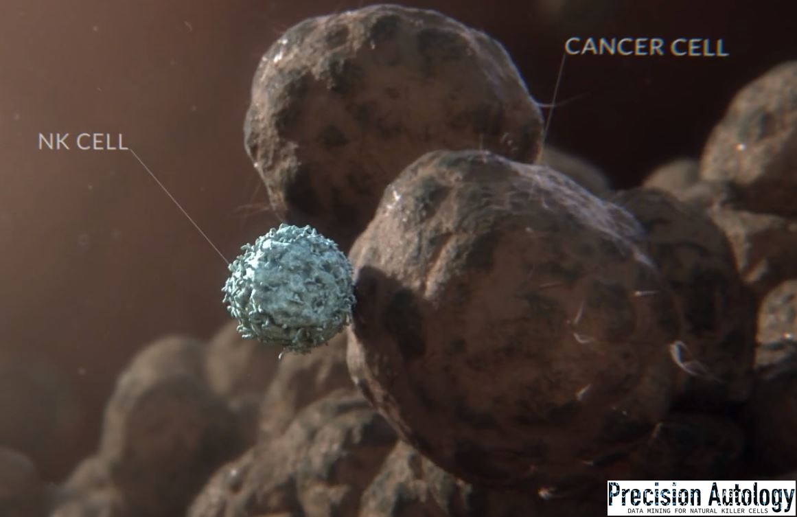
In a previous article I explained how allorecognition in Sea Squirts, from different colonies resembled polarization at the contact point of an immune synapse between a Natural Killer cell (NK) and its target.
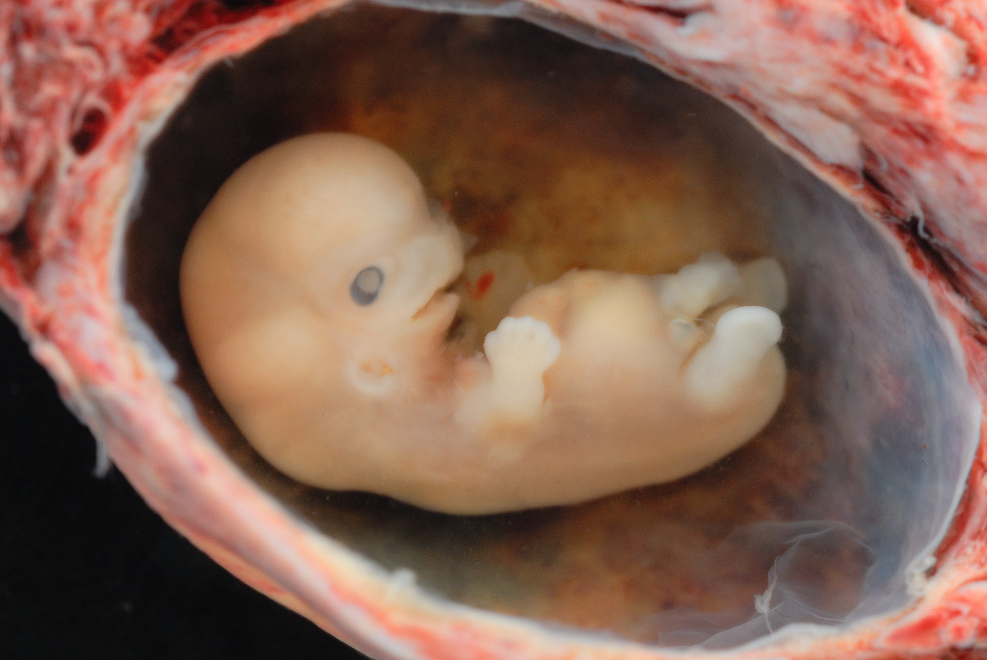
In early stage reproduction an “immunological paradox” occurs at the implantation site in the uterine wall where unexpected, male fertilized blastocyst is not rejected. Allorecognition occurs because maternal NK invades and remodels maternal blood vessels before the arrival of trophoblasts, the external cells of the blastocyst, that carry male antigens, during formation of the placenta that provides the fetus with nutrients.

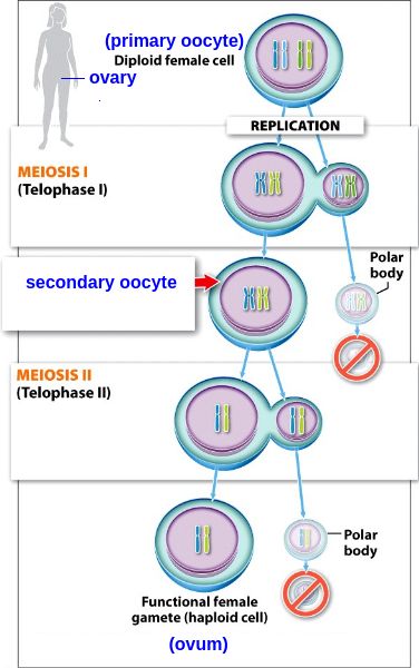
Sperm and ovum require gametes which are produced during meiotic cell division through phases of cellular polarization. Telophase (see image) resembles NK bound to its target just before the irreversible step that leads to perforin release and the target’s death. In this first Telophase, the yet to be separated pre-gametes contain twice the number of chromosomes, similarly when NK is enjoined to its target. Curiously, inhibitory and activating NK receptors that bind target cells diversify only through meiosis where in gametes the centrosome required for polarization has degenerated.

During meiosis as every gamete was made, research showed the first enzymatic step in meiotic recombination provoked a transient burst of p53 activity. Now it seems equally possible these p53 bursts could be attributed to relaxed transposon (mobile DNA) controls that accompany the meiotic process. Indeed, meiotic double-strand breaks are the proximal trigger for retrotransposon eruptions including in animals lacking p53.

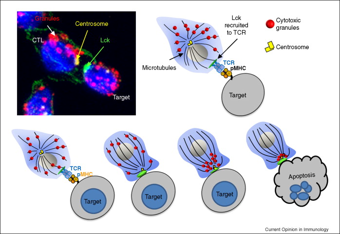
Mature NK can kill its target cell when combinations of inhibiting and activating receptors mediate a ‘non-self’ condition that triggers its polarization. At that point cytotoxic perforin and granzyme release toward the contact point, but NK can cancel the process. Reduced perforin release and binding in the immune synapse between these bound cells underlies the age-related decline in NK cytotoxicity. NK polarization and more generally cells undergoing division may also contribute to our understanding of ageing.

Courtesy
Here, we propose that certain features of the ageing process such as: (i) the increased reactivation rates of latent Mycobacterium tuberculosis, (ii) the slower resolution of inflammatory responses and (iii) the increased incidence of bacterial and fungal infection are attributable in part to an age-associated decline in NK function.

This paper demonstrated a pronounced age‐related impairment in perforin mobilization by NK to the K562 (target cell) contact point. It suggested that defective polarization to the immune synapse underlies the reduction in perforin secretion observed with age. The activation of mitogen‐activated protein kinases (MAPK) is critical for lytic granule (perforin-granzyme) polarization, granule exocytosis and NK Cytotoxicity. It is possible that these proximal signaling events are compromised by aging. In addition, it found that the studied p53 mutants regulated MAP2K3 gene whereas ectopic expression rescued the proliferative defect induced by mutant p53 knockdown.

Experimentally, NK that are absent of perforin or granzymes remain engaged five times longer. These locked-up NK cells would normally move to their next cell target to clear senescent or diseased cells, but cannot. NK suffering age-related reduced perforin release remain engaged awaiting a death signal from the target. This delay could be the single physiological cause of old or diseased cell accumulation in the body that is a primary cause of ageing. Perhaps polarization is causal?

Courtesy
Interestingly a member of the cytoskeleton superfamily Talin1 has been uniquely tied to two essential NK functions; 1. activation of LFA1, required for binding ICAM on NK target cell and, 2. NK polarization that results. We know overexpression of talin head activates LFA-1 and talin1 promotes cell proliferation by affecting the expression of BCL-2 family and p53 network. But, mdm2 the conserved nemesis of p53 is neutralized by Merlin, another cytoskeleton superfamily protein required for polarization. p53 also regulates the highly conserved Cdc42 which effects adhesion, actin cytoskeletal dynamics and cell movement including for angiogenesis in developing tumor microenvironments.

Through the p53 pathway Preimplantation Factor (PIF) is secreted by the blastocyst implant to reduce apoptosis in villous trophoblast lined vessels protruding as the developing placenta penetrates the decidua lining the uterine wall. Simultaneously maternal NK attack maternal endothelial cells lining arterial connections growing through the decidua. Opportunistically the apoptosis protected trophoblasts substitute dead maternal endothelial cells lining the connections. Thus mother succumbs to baby and the “immunological paradox” of maternal connection to embryonic placenta is overcome.

Furthermore, invalidation experiments demonstrate that PIF’s effects on placental apoptosis were mediated by TP53. Interestingly, the non-cancerous tissue associated with the over-expression of NK inhibiting ligand PCNA is the deciduas, and the only known NK cell subset that constitutively expresses NKp44 receptor for PCNA ligand is decidual NK cells. The PCNA gene is induced by p53 in the process of deciding cell fate, if PCNA is present in abundance in the absence of p53, DNA replication occurs.

However, fetal platelet antigens that cause miscarriages have been shown to prolong uterine/decidual NK survival, elevate NKp46 (LY94) receptor expression, perforin release and trophoblast apoptosis. The same receptor was also found to cluster at the NK-target cell immune synapse to regulate cytoskeletal organization and NK polarization on which perforin release is dependent.

Several common villous cells are implicated including NK, trophoblasts, vascular tip cells and cancer cells that may also induce NK to chaperone their effecting or replacing endo-epithelial cells lining host blood vessels. In the presence of senescent cells including NK with reduced perforin release, cancer cells may have discovered a trophoblast related back door to remodel blood vessels to serve transformed cells that ultimately grow to become cancerous tumors.

It’s distinctly possible that this p53 tug-o-war may be central to cellular age, death, reproduction and growth influenced by polarization and the perforin related effectiveness with which NK kills its meiosis evolved non-self targets.

At Precision Autology, the parent company of Codondex, we discover a patients p53 cell signatures to select their specific cancer cells, educate their natural killer cells to identify and kill the cancer that induced NK into its cooperative state.