Tuesday, October 10, 2023

Cancer's HLA-G Backdoor


piRNA actively control transposable elements (TE) that would otherwise disrupt genes, chromosomal stability, damage DNA, cause inflammation, disease and/or cell death. For example, increased levels of endogenous retroviruses (ERV), a TE subclass, trigger fibro inflammation and play a role in kidney disease development. However, in mammals, the transcription of TEs is important for maintaining early embryonic development. piRNA also function with TE's for important aspects of Natural Killer (NK) cell immune development. Regardless of the cell type, endogenous retroviral elements of the ERV1 family, are highly enriched at p53 sites highlighting the importance of this repeat family in shaping the transcriptional network of p53.

HLA/MHC are highly polymorphic molecules, expressed on cells and recognized by NK cells. In mammals it is necessary to generate specialized NK cell subsets that are able to sense changes in the expression of each particular HLA molecule.

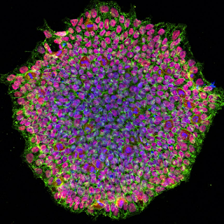
Decidual natural killer cells (dNK), the largest population of leukocytes at the maternal–fetal interface, have low cytotoxicity. They are believed to facilitate invasion of fetal HLA-G+ extravillous trophoblasts (EVT) into maternal tissues, essential for establishment of healthy pregnancies. dNK interaction with EVT leads to trogocytosis that acquires and internalizes HLA-G of EVT. dNK surface HLA-G was reacquired by incubation with EVT's. Activation of dNK by cytokines and/or viral products resulted in the disappearance of internalized HLA-G and restoration of cytotoxicity. Thus, the cycle provides both for NK tolerance and antiviral immune function by dNK.

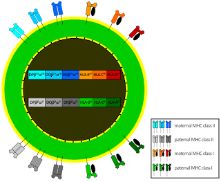
A remote enhancer L, essential for HLA-G expression in EVT, describes the basis for its selective  immune tolerance at the maternal–fetal interface. Found only in genomes that lack a functional HLA-G classical promoter it raises the possibility that a retroviral element was co-opted during evolution to function in trophoblast-specific tolerogenic HLA/MHC expression. CEBP and GATA regulate EVT expression of HLA-G through enhancer L isoforms.

HLA-G1 is acquired by NK cells from tumor cells, within minutes, by activated, but not resting NK cells via trogocytosis. Once acquired, NK cells stop proliferating, are no longer cytotoxic and behave as suppressors of cytotoxic functions in nearby NK cells via the NK ILT2 (Mir-7) receptor. Mir-7 is a well researched intervention target in inflammatory diseases and belongs to a p53-dependent non-coding RNA network and MYC signaling circuit.

Cells that transcribe enhancer L isoforms and HLA-G, feed NK cells with HLA-G as an innate element for self determination, similar to the way EVT's restrain cytotoxicity of dNK. Then incoming, NK cells at the periphery of tumor microenvironments (TME) may promote vascular remodeling, as in the uterus during pregnancy, by acidifying the extracellular matrix with a2V that releases bound pro-angiogenic growth factors trapped in the extracellular matrix. After that these incoming NK cells succumb to the influence of Mir-7 resulting in low cytotoxic, inactive NK in the TME. 

Discovering resistant NK cells in the TME of a patient, for incubation, expansion and activation is a Codondex precision therapy objective based on p53 computations.


Wednesday, September 27, 2023

When Immunity Fails Programmed Cell Death

DNA Damage Response

Telomeric repeat (TR) sequences are responsible for genome integrity, where instability is a primary factor that leads to activation of p53. Introduction of a TR into cells leads to stabilization of p53, specific to TRs and not observed in plasmids containing non-TR sequences. TR-activated p53 exhibited enhanced transcriptional activity and induced p53-dependent growth suppression, measured as a reduction in colony formation. Sub-telomeric p53 binding prevents accumulation of DNA damage at human telomeres.  

Healthy cells experience thousands of DNA lesions per day. Micronuclei, containing broken fragments of DNA or chromosomes, that have become isolated, are recognized as one mediator of DNA damage response (DDR)-associated immune recognition. Like micronuclear DNA, mitochondrial DNA (mtDNA) is recognized by cGAS to drive STING-mediated inflammatory signaling. Mitochondrial damage can intersect DNA repair and inflammatory cascades with programmed cell death, through p53. In human fibroblasts and conditionally immortalized vascular smooth muscle cells p53 mediates CD54 (ICAM-1) overexpression in senescence.

Replicative senescence, an autophagy dependent program and crisis are anti-proliferative barriers that human cells must evade to gain immortality. Telomere-to-mitochondria signaling by ZBP1 mediates replicative crisis. Dysfunctional telomeres activate innate immune responses (IFN) through mitochondrial TR RNA (TERRA)–ZBP1 complexes. Senescence occurs when shortened telomeres elicit a p53 and RB dependent DNA-damage response. A crisis-associated isoform of ZBP1(innate immune sensor) is induced by the cGAS–STING DNA-sensing pathway, but reaches full activation only when associated with TERRA transcripts from dysfunctional telomeres. p53 utilizes the cGAS/STING innate immune system pathway for both cell intrinsic and cell extrinsic tumor suppressor activities. cGAS-STING activation induces the production of IFN-b and increases CD54 expression in  human cerebral microvascular endothelial cells.

In melanoma patients there is a significant correlation between cGAS expression levels and survival and between NK cell receptor expression levels and survival. Loss of cGAS expression by tumor cells could permit the tumor cell to circumvent senescence or prevent immunostimulatory NKG2D ligands expression. Loss of p53 and gain of oncogenic RAS exacerbated pro-malignant paracrine signaling activities of senescence-associated secretory phenotypes. Results imply that heterogeneity in cGAS activity, across tumors, could be an important predictor of cancer prognosis and response to treatment and suggest that NK cells could play an important role in mediating anti-tumor effects. Coculture of wild-type p53-induced human tumor cells with primary human NK cells enhanced NKG2D-dependent degranulation and IFN-γ production by NK cells. 

When p53 consensus sequences are modified and DNA damage response is compromised, replicative crisis ensues, mitochondrial membranes misfunction, mtDNA expression is downregulated and IFN signaling upregulates. A cell may then express activating immune ligands that bind NK receptors signaling non-self and cytolytic death or inhibitory receptors that signal self and immortality


Thursday, September 21, 2023

Indispensable Mitochondria - Cancers back door?


Immediately prior to fertilization spermatozoa are devoid of Mitochondrial DNA (mtDNA), potentially explaining an aspect about selection that may serve the legacy for maternal immune tolerance. Post fertilization, on day 11-13, outermost trophoblasts of the blastocyst dock with the decidual lining as it embeds in the uterine wall. Then, maternal vascular remodeling and placental formation begin toward successful implantation. 

Higher quality trophoblasts are associated with lower mtDNA content. Moreover, euploid blastocysts with higher mtDNA content had a lower chance to implant and mtDNA replication is strictly downregulated between fertilization and the implantation. What is it about absent or reduced mtDNA that may also relate to the mechanics of immune tolerance and vascular remodeling, which are also features of solid tumors.

The initial absence or downregulation of MtDNA, may relate an immune tolerance by uterine Natural Killer (NK) cells. As mtDNA upregulates, after day 12, it may initiate NK auto-reactivity required for maternal microvascular remodeling. This auto-immune paradox is a prerequisite for vascular remodeling, which is also seen in localized hypertension, and the likely basis of successful blastocyst implantation. Acutely, micro-hypertension induced mechanical stretch, on endothelial cells, interconnects innate and adaptive immune responses. 

The dominant cell in the decidua is an NK subset (dNK), they express low levels of IFN-γ and express proteins of Renin Angiotensin System (RAS). At day 12 RAS peptide ANP colocalizes to dNK’s suggesting that dNK RAS infers localized responsiveness.  When TFAM, required for transcription of mtDNA, was deleted from cardiomyocytes, after 32 days, animals developed cardiomyopathy and Nppa (gene encoding ANP) and Nppb expression was elevated. 

In monocytes increased endothelial stretch activates STAT3, which is involved in driving almost all pathways that control NK cytolytic activity and reciprocal regulatory interactions between NK cells and other components of the immune system. The crosstalk between STAT3 and p53/RAS signaling controls cancer cell metastasis. p53, Stat3, and, potentially, the estrogen receptor are thought to act as co-regulators, affecting mitochondrial gene expression through protein-protein interactions. Co-immunoprecipitation of p53 with TFAM suggests it may regulate mitochondrial DNA-damage repair.

Like initial trophoblasts with low level mtDNA, mature cells, like cardiomyocytes that prolong low level mtDNA may also aggravate autoimmune sponsored hypertension that remodels microvascular networks providing nutrients for growth of reduced mtDNA stem cell replicas. Indeed, mitochondrial dysfunction (from depleted mtDNA) does not affect pluripotent gene expression, but results in severe defects in lineage differentiation.

During severe sepsis, intense, on-going mtDNA damage and mitochondrial dysfunction could overwhelm the capacity for mitochondrial biogenesis, leading to a gradual decline in mtDNA levels over time. This may contribute to monocyte immune deactivation, which is associated with adverse clinical outcomes and could be reversed by IFN-γ

Identifying cells that optimally educate cocultured NK cells for precision IFN-γ and cytolytic responsiveness is part of the ongoing work by the Codondex team.


Saturday, August 19, 2023

Can Ancient Pathways Defeat Cancer?


It has been widely acknowledged that non-coding RNAs are master-regulators of genomic function. The association between human introns and ncRNAs has a pronounced synergistic effect with important implications for fine-tuning gene expression patterns across the entire genome. There is also strong preference of ncRNA from intronic regions particularly associated with the transcribed strand. 

Accumulating evidence demonstrates that, analogous to other small ncRNAs (e.g. miRNAs, siRNA's etc.) piRNAs have both oncogenic and tumor suppressive roles in cancer development. Functionally, piRNAs maintain genomic integrity and cell age by silencing repetitive, transposable elements, and are capable of regulating the expression of specific downstream target genes in a post-transcriptional manner. 

Unlike miRNAs and siRNAs, the precursors of piRNAs are single stranded transcripts without any prominent secondary hairpin structures. These precursors are usually generated from specific genomic locations containing repetitive elements, a process that is typically orchestrated via a Dicer-independent pathway. 

Without restraint, the ancient, L1 class of transposable elements can interrupt the genome through insertions, deletions, rearrangements, and copy number variations. L1 activity has contributed to instability and evolution of genomes, and is tightly regulated by DNA methylation, histone modifications, and piRNA. They can impact genome variation by mispairing and unequal crossing-over during meiosis due to repetitive DNA sequences. Indeed meiotic double-strand breaks are the proximal trigger for retrotransposon eruptions as highlighted in animals lacking p53.

Through a novel 28-base small piRNA of the KIR3DL1 gene, antisense transcripts mediate Killer Ig-like receptor (KIR) transcriptional silencing in immune somatic, Natural Killer (NK) cell lineage, a mechanism that may be broadly used in orchestrating immune development. Expressed on NK cells, KIR's are important determinants of NK cell function. Silencing  individual KIR genes is strongly correlated with the presence of CpG dinucleotide methylation within the promoter. 

Structural research exposed the enormous binding complexity behind KIR haplotypes and HLA allotypes. Not only via protein structures, but also plasticity and selective binding behavior's as influenced by extrinsic factors. One study links a specific recognition of HLA-C*05:01 by KIR2DS4 receptor through a peptide highly conserved among bacteria pathogenic in humans. Another demonstrated a hierarchy of functional peptide selectivity by KIR–HLA-C interactions, including cross-reactive binding, with relevance to NK cell biology and human disease associations. Additionally a p53 peptide most overlapped other high performance peptides for a HLA-C allotype C*02:02 that shares identical contact residues with C*05:01.

Ancient pathways linking p53 to attenuation of aberrant stem cell proliferation may predate the divergence between vertebrates and invertebrates. Human stem cell proliferation, as determined by p53 transposable element silencing, may also serve a NK progenitor to promote the repertoire of more than 30,000 NK cell subsets

A recent study showed that wild type p53 can restrain transposon mobility through interaction with PIWI-piRNA complex. Also, cellular metabolism regulates sensitivity to NK cells depending on P53 status and P53 pathway is coupled to NK cell maturation leaving open the possibility that a direct relationship exists. Further, functional interactions between KIR and HLA modify risks of basal cell carcinoma (BCC) and squamous cell carcinomas (SCC) and KIR B haplotypes provide selective pressure for altered P53 in BCC tumors

Anticipating p53's broader influences or responses, cells, extracted from 48 different sections of 7 tumor biopsies were sequenced and TP53 DNA computed using Codondex algorithm. Each section produced a TP53 Consensus Variant (CV), represented by its intron1, ncDNA Key Sequence's (KS). Bioinformatic correlations between each KS and cytotoxicity resulting from NK coculture with the section may predict KIR-HLA and extrinsic factor plasticity to reliably determine from KS's, optimal cell/tissue selections for NK cell education and licensing. 


Wednesday, May 17, 2023

Immune Synchronization

Stem Cell

Navigating the regulatory regimes that govern drug safety can be challenging. But, rigorous standards are more relaxed in the lesser used track for autologous and/or minimally manipulated cell treatments. Toward meeting the challenges of this minimal regulation track, the wide-spectrum of NK cells, of the innate immune system, are compelling candidates to address complex cellular and tissue personalization's or conditions of disease. One effect of cell function on NK cell potency occurs via aryl hydrocarbon receptor (AhR) dietary ligands, potentially explaining numerous associations that have been observed in the past.

The AhR was first identified to bind the xenobiotic compound dioxin, environmental contaminants and toxins in addition to a variety of natural exogenous (e.g., dietary) or endogenous ligands and expression of AhR is also induced by cytokine stimulation. Activation with an endogenous tryptophan derivative, potentiates NK cell IFN-γ production and cytolytic activity which, in vivo, enhances NK cell control of tumors in an NK cell and AhR-dependent manner.

A combination of ex vivo and in vivo studies revealed that Acute Myeloid Leukemia (AML) skewed Innate Lymphoid Cell (ILC) Progenitor towards ILC1's and away from NK cells as a major mechanism of ILC1 generation. This process was driven by AML-mediated activation of AhR, a key transcription factor in ILC's, as inhibition of AhR led to decreased numbers of ILC1's and increased NK cells in the presence of AML.

Activation of AhR also induces chemoresistance and facilitates the growth, maintenance, and production of long-lived secondary mammospheres, from primary progenitor cells. AhR supports the proliferation, invasion, metastasis, and survival of the Cancer Stem Cells (CSC's) in choriocarcinoma, hepatocellular carcinoma, oral squamous carcinoma, and breast cancers leading to therapy failure and tumor recurrence.

Loss of AhR increases tumorigenesis in p53-deficient mice and activation of p53 in human and murine cells, by DNA-damaging agents, differentially regulates AhR levels. Activation of the AhR/CYP1A1 pathway induces epigenetic repression of many tumor suppressor and tumor activating genes, through modulation of their DNA methylation, histone acetylation/deacetylation, and the expression of several miRNAs. 

p53 is barely detectable under normal conditions, but levels begin to elevate and locations change particularly in cells undergoing DNA damage. The significant network effect of p53 availability and its mutational status in cancer makes it the worlds most widely studied gene. 

From 48 sequenced samples of two different tumors, Codondex identified 316 unique Key Sequences (KS) of the TP53 Consensus. 9 of these contained the core AhR 5′-GCGTG-3′ binding sequence, and some overlapped p53 quarter binding sites as illustrated below;

Key Sequence                                                                           

GGATAGGAGTTCCAGACCAGCGTGGCCA (intron1) AhR [1699,1726], p53 @ [1706,1710]

AAAAATTAGCTGGGCGTGGTGGGTGCCT (intron1) AhR [1760,1787], p53 [1783,1787]

AAAAAAAATTAGCCGGGCGTGGTGCTGG (intron6) AhR [12143,12170]

GAGGCTGAGGAAGGAGAATGGCGTGAAC (intron6) AhR [12195,12222]

We propose that DNA damage liberates transposable DNA elements that are normally repressed by p53 and other suppressor genes. The p53 repair/response also includes increased cooperation between p53 and AhR, which further influence transcription, mRNA splicing or post-translation events. Repeated damage, at multi-cellular scale, may proximally bias ILC's toward NK cells capable of specific non-self detection, through localized ligand, receptor relationships that trigger cytolysis and immune cascades. 

KS's are a retrospective view of transcripts ncDNA elements, ranked by cDNA that may reflect inherent bias that can be used to direct NK cell education. One way to accomplish minimal manipulation may be to leverage patient immunity by educating autologous NK cells with computationally selected tumor cells, identified by KS alignments to the index of past experiments that expanded and triggered a more desirable immune response. Customizable immune cascades, capable of managing disease or preventatively supporting a desired heterogeneity being the primary objective. 


Tuesday, March 21, 2023

Tolerating Your Non-self!

Immune cells get comfortable with cancer
Courtesy https://deepai.org

A hallmark of cancer, autoimmunity and disease is the aberrant transcription of typically silenced, repetitive genetic elements that mimic Pathogen-Associated Molecular Patterns (PAMP's) that bind Pattern Recognition Receptors (PPR's) triggering the innate immune system and inflammation. Unrestrained, this 'viral mimicry' activates a generally conserved mechanism that, under restraint, supports homeostasis. These repetitive viral DNA sequences normally act as a quality control over genomic dysregulation responding in ways that preferentially promote immune conditions for stability. If aberrantly unrestrained and the 'viral mimicry' is transcribed it may result in undesirable immune reactions that disrupt the homeostasis of cells.

Mitochondrial DNA (mtDNA) are one source of cytosolic double stranded RNA (dsRNA) that is commonly present in cells. Trp53 Mutant Embryonic Fibroblasts (MEF's) contain innate immune stimulating endogenous dsRNA, from mtDNA that mimic PAMP's. The immune response, via RIG-1 like PRR, leads to expression of type 1 interferon (IFN) and proinflammatory cytokine genes. Further, Natural Killer cells also produce a multitude of cytokines that can promote or dampen an immune response. Wild-type p53 suppresses viral repeats and contributes to innate immunity by enhancing IFN-dependent antiviral activity independent of its function as a proapoptotic and tumor suppressor gene. 

Post-translationally modified P53, located in the cytoplasm, enhances the permeability of the mitochondrial outer membrane thus stimulating apoptosis. However, treating Trp53 mutant MEF's with DNA demethylating agent caused a huge increase in the level of transcripts encoding short interspersed nuclear elements and other species of noncoding RNAs that generated a strong type 1 IFN response. This did not occur in p53 wild-type MEF's. Thus it appears that another function of p53 is to silence repeats that can accidentally induce an immune response.

This has several implications for how we understand self versus non-self discrimination. When pathogen-associated features were quantified, specific repeats in the genome not only display PAMP's capable of stimulating PRRs but, in some instances, have seemingly maintained such features under selection. For organisms with a high degree of epigenetic regulation and chromosomal organization immuno-stimulatory repeats release a danger signal, such as repeats released after p53 mutations. Here, immune stimulation may act as back-up for the failure of other p53 functions such as apoptosis or senescence due to mutation. This supports the hypothesis that specific repeats gained favor by maintaining non-self PAMPs to act as sensors for loss of heterochromatin as an epigenetic checkpoint of quality control that avoids genome instability generally. 

When P53 mutates it begins to fail its restraint of viral suppression, this enables a 'viral mimicry' and aberrant immune reactions. These may promote survival of cells that can leverage immunity, promote angiogenesis and heightened proliferation of cancers, or other diseases under modified conditions for non-self tolerance.