Showing posts with label mhc. Show all posts

Saturday, August 8, 2026

Do p53 Repeat Fields Synchronize Target Cells and Natural Killer Cells?


Natural Killer cells face a remarkable biological problem. They must continuously distinguish healthy cells from stressed, infected or malignant cells and decide which should be destroyed. Increasing evidence suggests that p53 participates directly in coordinating that decision. A 2024 review in Frontiers in Immunology brings together evidence that p53 affects not only the internal fate and external immune visibility of the target cell, but also the homeostasis, receptor signalling and functional state of the Natural Killer cell itself.

This creates an intriguing question for Codondex. If p53 coordinates several different components of the NK-target interaction, could the unusually dense repeat structures that Codondex detects within TP53 help identify genomic regions involved in regulating that coordination? We describe these structures as High-Density Nested Repeat Fields, or HDNRFs: regions in which repeated, overlapping and nested short sequences become unusually concentrated. HDNRFs are not simply conventional tandem repeats. They represent a broader sequence geography that becomes visible when DNA is interrogated simultaneously across different sequence lengths and positions.

The Codondex methodology approaches this problem without first needing to know the biological function of the sequence. It divides genetic sequence into overlapping components, preserves their relative positions and relationships, and identifies sequence combinations that recur or associate unusually strongly. The important distinction is that Codondex can identify unusual sequence architecture first and ask what that architecture means biologically afterwards. The progression is therefore from sequence, to pattern, to biological association and finally, if experimentally demonstrated, to mechanism.

We have already seen an intriguing example of this process within TP53. Earlier Codondex analysis converged on the intron 3 region containing the known PIN3 polymorphism, or Polymorphism Intron 3. PIN3 is a 16-base-pair duplication situated within an unusual region of TP53 associated with structural and regulatory features affecting p53 RNA processing. The importance of the PIN3 observation is not that Codondex discovered PIN3 itself. The polymorphism was already known. What was interesting was the route by which Codondex arrived there. The algorithm was not instructed to search for PIN3 or for a known p53 regulatory region. It converged on the location because of an unusual concentration of relationships within the sequence itself. Conventional biology then provided an independent reason why that location could matter.


PIN3 therefore provided an early example of the utility we are now trying to test more systematically with HDNRFs: whether unusual repeat geography can lead to biologically significant regulatory locations before we understand why those locations are important. More recent Codondex analysis has strengthened that question by revealing concentrations of repeated and overlapping sequences elsewhere within TP53 and other genes, suggesting that repeat density may represent a genomic characteristic worth measuring in its own right.

A second observation made the p53 question considerably more interesting. In our previous analysis of 48 tumour sections, Codondex-derived sequence rankings were compared with several experimental immune measurements, including Chromium-release Natural Killer cytotoxicity under unstimulated pNK, IL-2-stimulated pNK and sNK conditions, together with IFN-γ and other measurements. When the sequences were examined in different ways, including duplicate occurrence, repeated short sequences and tandem or overlapping repeat relationships. pNK repeatedly emerged as one of the more consistent candidate associations.

This was unexpected because the unstimulated pNK experimental signals themselves were frequently weak. Many tumour sections showed little or no measured pNK activity, while stimulated NK measurements often generated much stronger absolute signals. Yet pNK continued to appear when independent measures of Codondex sequence structure were compared with the experimental results. We previously discussed the possibility that this recurrence may therefore be more informative than the absolute strength of the pNK measurement suggests. A weak biological signal that repeatedly aligns with independently derived sequence characteristics may be pointing to an underlying state rather than simply reflecting the magnitude of immune activity.

The Frontiers in Immunology review provides a biological framework capable of explaining why this might occur. In the target cell, wild-type p53 can increase expression of NK-activating ligands including ULBP1 and ULBP2, which interact with NKG2D, and can influence PVR/CD155 and other components of activating versus inhibitory NK recognition. p53 also participates in the apoptotic machinery determining whether an NK attack successfully kills its target, including BAX-dependent mitochondrial pathways and death-receptor mechanisms.

But importantly, p53 is not operating only within the target. The same review describes p53-related regulation within NK cells themselves. p53 participates in NK-cell cycle and apoptotic control, influences receptor expression and regulates SAP, which couples SLAM-family signalling through Fyn and Vav-1 to NK activation. The authors explicitly separate the regulatory role of p53 in NK cells from its role in tumour cells, and conclude that these functions contribute to NK recognition and targeting of malignant cells.

This means that p53 potentially operates on both sides of the immune synapse. In the target, it can contribute to making a damaged cell visible, engageable and susceptible to killing. In the NK cell, p53-related pathways can influence whether the immune cell is functionally competent to recognize and respond to that target. Successful cytotoxicity may therefore depend upon the state of two interacting cellular programmes becoming appropriately aligned.

We have described this concept as p53-driven NK-target synchronicity. Until recently, however, the word “synchronicity” was principally a functional description of several p53-dependent variables moving together. New experimental work published in Molecular Systems Biology now gives that term a much more literal biological foundation.

Venkatachalapathy and colleagues demonstrated that p53 behaves as a dynamic cellular oscillator whose normally heterogeneous pulses can be experimentally phase-reset and synchronized across individual cells. Using time-lapse microscopy and controlled DNA-damage stimulation in MCF-7 cells, they showed that p53 oscillations could be brought into significantly greater temporal alignment by appropriately timed repeated stimuli. Two damage pulses spaced approximately 4.0 to 5.5 hours apart produced significantly increased synchronization compared with a single stimulus.

This is important because the synchronization was not merely cosmetic. Altering p53 pulse timing changed downstream gene-expression dynamics, and phase resetting affected cellular fate. The study showed that changes in p53 oscillatory frequency altered expression of p53 target genes and that greater synchronization reduced the probability that cells escaped cell-cycle arrest. In other words, p53 synchronization produced synchronized biological consequences.

This materially strengthens the hypothesis we have been developing. p53 should no longer be viewed only as a molecular switch whose biological significance is determined by how much p53 is present. Its timing, frequency and phase also contain biological information. The new work demonstrates experimentally that p53 dynamics can be reset, that cells can be brought into temporal alignment, and that changing those dynamics changes downstream molecular programmes and cell fate.

The implication for Codondex is potentially significant. A p53 HDNRF need not simply influence whether there is “more” or “less” p53. If repeat architecture has regulatory significance, it could conceivably affect p53 expression, RNA processing, response thresholds, pulse amplitude, oscillatory frequency or the persistence of downstream signals. PIN3 becomes particularly interesting in this context because Codondex independently converged on an intronic TP53 region already associated with regulation of TP53 processing. The question may therefore be broader than whether repeat fields alter p53 abundance. It may be whether they identify or influence the dynamic state of the p53 regulatory system.

This offers a new way of interpreting the repeated pNK result. Unstimulated pNK cytotoxicity may represent a relatively delicate basal relationship between the target cell and the Natural Killer cell. For killing to occur, the target must become sufficiently visible, activating signals must overcome inhibitory signals, the NK cell must be in a competent functional state, the two cells must successfully engage and the target must remain susceptible to the resulting cytotoxic attack. These conditions need not individually produce large experimental signals. Their biological importance may lie in whether they occur together.

The emerging hypothesis is therefore that the Codondex repeat signal is associated not simply with “NK activity,” but with the state in which the target and NK cell become appropriately coordinated. The target-side p53 programme can influence recognition ligands and apoptotic susceptibility, while the NK-side p53 programme can influence cellular homeostasis and activation pathways. The new Molecular Systems Biology work adds the crucial observation that p53 itself possesses a genuine temporal synchronization mechanism capable of coordinating downstream biological responses.

It is important not to claim more than the evidence demonstrates. The new study did not place an NK cell beside a tumour cell and show that p53 oscillations in the two cells phase-lock to one another. It synchronized p53 oscillations across MCF-7 cells using externally delivered DNA-damage pulses. It therefore proves that p53 is a synchronizable biological oscillator, but does not yet prove that NK and target-cell p53 oscillators synchronize during immune surveillance. That distinction defines the next experiment rather than weakening the hypothesis.

The research question can now be framed much more precisely. Tumour targets should be classified according to their relevant Codondex HDNRF state and then observed together with Natural Killer cells while p53 dynamics are measured in real time. Instead of measuring p53 only at a single time point, the experiment should measure p53 pulse timing, frequency, amplitude and phase in the target and, where technically possible, in the NK cell. These measurements should be followed simultaneously by target-cell ULBP1/2, PVR/CD155 and other recognition signals, NK activation and engagement, CD107a degranulation, IFN-γ production and ultimately target-cell killing.

The decisive observation would be whether these variables move together in time. Does a change in target-cell p53 state precede altered NK-recognition ligand expression? Does successful NK engagement occur preferentially during a particular target-cell p53 state? Does the NK cell itself undergo a corresponding p53-dependent state change? Do successful killing events show greater coordination between these trajectories than unsuccessful encounters? And most importantly for Codondex, does the strength or architecture of the relevant HDNRF predict any of these dynamic relationships?

The experimental design can then move from association to causality. Removing or inhibiting functional p53 should disturb the predicted relationship. Restoring p53 should restore at least part of it. Directly altering a candidate HDNRF using locus-specific sequence or epigenetic intervention could then determine whether the repeat field is merely a marker of the p53 state or participates in establishing it. If changing the HDNRF changes p53 dynamics and those changes propagate through NK recognition and cytotoxicity, the evidence would move substantially closer to a causal genomic mechanism.

PIN3, the recurring pNK association and the new p53 synchronization data now form three independent but potentially connected observations. PIN3 showed that Codondex could converge computationally on an unusual TP53 sequence region already recognized by conventional biology as regulatory. The tumour-section analysis repeatedly brought pNK to the surface despite weak absolute unstimulated NK signals. The new phase-resetting experiments demonstrate that p53 itself can operate as a synchronizable dynamic system whose temporal state changes downstream gene expression and cellular fate.

None of these observations alone proves that an HDNRF synchronizes a Natural Killer cell with its target. Together, however, they produce a considerably more specific and experimentally falsifiable hypothesis. A p53 repeat field may identify or influence a dynamic genomic state that helps coordinate p53-dependent programmes in the target and Natural Killer cell, increasing the probability that recognition, engagement, susceptibility and cytotoxic response occur together.

That hypothesis also provides a possible explanation for why pNK keeps appearing in the Codondex analysis. The algorithm may not be detecting the strength of an immune response. It may instead be detecting sequence architecture associated with the underlying state that permits natural NK surveillance to occur.

Codondex was designed to find relationships in genetic sequence before we necessarily know what those relationships mean. PIN3 was an early indication that concentrated sequence relationships could converge on known p53 regulatory biology. The recurring pNK evidence suggested a possible connection with Natural Killer surveillance. We now know experimentally that p53 itself is capable of phase resetting and synchronization, and that synchronized p53 dynamics can alter downstream biological outcomes.

The next question is therefore unusually clear: do p53 repeat fields participate in setting the dynamic conditions under which a target cell and a Natural Killer cell become synchronized for recognition and killing?

That is now an experiment worth doing.


Thursday, March 26, 2026

When Processing, Not Presence, Determines Visibility


It is easy to assume that if a protein accumulates in a diseased cell, the immune system will eventually see it. In the case of p53, that assumption has always had an intuitive appeal. p53 is one of the central stress-response proteins in biology, frequently altered in cancer, often stabilized, and deeply woven into the molecular logic of cell fate. If any intracellular protein should become immunologically visible, it ought to be p53.

But the deeper one looks at antigen presentation, the less that simple view holds. What matters is not merely whether p53 is present. What matters is whether peptide fragments derived from p53 are generated in the right form, survive intracellular trimming, fit the preferences of a particular HLA groove, and remain stable enough on the cell surface to be interrogated by either a T cell or an NK-cell receptor system. The 2022 Codondex article, Expanding Treatment Horizons, was already moving in that direction by highlighting an underappreciated observation from the HLA-C ligandome literature: a TP53-derived peptide, TAKSVTCTY, was identified as a naturally presented ligand of HLA-C*02:02. That observation comes from Moreno Di Marco and colleagues’ immuno-peptidomics study, which also listed MAGEA3-derived peptides among ligands presented by the same allotype.

That point remains important, but it also needs sharpening. The HLA-C paper tells us that a TP53-derived peptide can be naturally presented by HLA-C02:02. It does not tell us that HLA-C02:02 is already a dominant or clinically validated p53 presentation route in the way that HLA-A02:01 has become. For that, the literature is far stronger on the HLA-A side. A substantial body of work has shown that **wild-type p53 peptides presented by HLA-A02:01**, especially the well-known p53(264–272) epitope LLGRNSFEV, can stimulate cytotoxic T-cell responses and can be recognized on tumor cells. This was shown in studies such as Chikamatsu et al. Hoffmann et al. Gnjatic et al. and later vaccine-oriented work including Svane et al and the broader review literature on p53-targeting vaccines. In other words, for HLA-A*02:01, p53 is not just a theoretical ligand source; it is already part of a fairly mature immunotherapeutic story.

The most useful contribution of the recent Nature paper, The DNA virome varies with human genes and environments, is that it sharpens the mechanistic frame through which both HLA-C02:02 and HLA-A02:01 should now be viewed. The paper is not a p53 paper. It does not center tumor antigens, and it does not establish anything directly about TP53 peptide presentation. What it does show, at population scale, is that viral DNA load is shaped not only by HLA variation but also by the antigen-processing machinery, especially ERAP1 and ERAP2. That matters because it shifts the center of gravity away from a simplistic “does the peptide bind?” model and toward a more realistic “does the peptide survive the whole processing pipeline?” model.

That shift is especially important for p53. The HLA-A02:01 literature had already hinted that presentation of the classic p53(264–272) epitope depends on more than sequence alone. The work by Kuckelkorn et al showed that generation of this epitope is influenced by the interferon-γ-inducible processing machinery and that a hotspot mutation at residue 273 can prevent proper generation of the epitope. This is a reminder that even for the most familiar p53/HLA-A02:01 peptide, presentation is a processing problem before it becomes a recognition problem. The Nature virome study widens that principle: inherited variation in antigen processing can have measurable biological consequences at human scale. Read together, these papers suggest that p53 visibility is governed not simply by the existence of a fitting sequence, but by whether intracellular processing delivers that sequence intact to the appropriate HLA molecule.

This is where the contrast between HLA-A02:01 and HLA-C02:02 becomes genuinely interesting. HLA-A02:01 has a long experimental trail behind it: peptides were mapped, CTLs were induced, tumors were shown to present certain epitopes, and vaccine studies were built on top of that scaffold. HLA-C02:02, by contrast, remains more conditional. The ligandome study establishes that TAKSVTCTY from TP53 can indeed appear on HLA-C02:02, and it also gives a broader view of the peptide preferences of that allotype. In that same work, HLA-C02:02 is described as favoring small aliphatic or hydrophilic residues at position 2, with additional motif features helping define its ligand space. That does not diminish the importance of the TP53 observation; it means the TP53 peptide should be treated as a real but selective presentation event rather than assumed to be broadly immunodominant.

The biology becomes even more layered because HLA-C is not simply a lower-profile version of HLA-A. HLA-C occupies a distinct place in immune regulation. Compared with HLA-A and HLA-B, HLA-C is generally expressed at lower surface levels and is more tightly integrated with KIR-mediated NK-cell regulation. That broader point is well summarized in the Nature Communications paper Structural and regulatory diversity shape HLA-C protein expression levels, which notes both the lower surface expression of HLA-C and its extensive functional relationship with KIRs. This makes HLA-C particularly interesting for p53 because a peptide displayed by HLA-C is not only a possible T-cell target; it is also part of a signaling surface read by NK cells.

That NK dimension turns out not to be merely background context. More recent work has shown that KIR recognition of HLA-C is often peptide-dependent. The point is made clearly in studies such as Sim et al. 2017 and Sim et al. 2023: the HLA-C molecule is not being read in a peptide-blind way. Inhibitory and activating KIRs can be strongly shaped by the identity of the peptide bound in the groove. That has profound implications for any discussion of TP53 peptides on HLA-C02:02. A TP53-derived peptide on HLA-C02:02 may not simply mark a cell for CD8 T-cell inspection; it may also alter the threshold for NK inhibition or activation. This is one of the most important places where the older Codondex article and the newer immunogenetic literature genuinely converge.

So the corrected reading is not that the 2026 Nature paper newly proves something specific about HLA-C*02:02 presenting p53. It does not. What it does is make the older HLA-C02:02 observation more meaningful by placing it inside a stronger mechanistic framework. The question is no longer only whether TAKSVTCTY can bind HLA-C02:02; the question is whether an individual’s processing machinery, inflammatory state, and HLA context allow that peptide to be generated, preserved, loaded, displayed, and then interpreted by either T cells or NK cells in a biologically consequential way. That is a more demanding question, but it is also a more interesting one.

This also helps explain why HLA-A*02:01 remains the more established p53 route. The A02:01 pathway has yielded peptides that are repeatedly recoverable in experimental systems, repeatedly recognized by CTLs, and repeatedly leveraged in translational work. The HLA-C02:02 pathway looks more contingent: real, but likely more dependent on peptide selection pressure, trimming, and the NK-facing consequences of peptide-loaded HLA-C. Seen this way, HLA-A02:01 is the clearer adaptive pathway, while HLA-C02:02 may be a narrower but potentially more intriguing bridge between tumor antigen presentation and innate immune tuning.

That may be the most useful lesson from putting these papers together. p53 is not simply “presented” or “not presented.” It passes through a filter. In HLA-A02:01, that filter has already produced a clinically legible signal. In HLA-C02:02, the signal is fainter, but perhaps more information-rich, because it may be read simultaneously by T cells and NK-cell receptor systems. If that is right, then the next real step is not more speculation about binding motifs alone. It is experimental work that directly compares TP53 peptide generation, ERAP dependence, surface abundance, and KIR/TCR consequences across HLA-A02:01 and HLA-C02:02 backgrounds. That is where the overlap becomes testable rather than merely suggestive.

Tuesday, March 3, 2026

Natural Killers, Mitochondria, p53, and Parkinson’s


The emerging landscape of neuro-immune communication reveals that the traditional boundaries between immune sentinel function and neuronal integrity are far less distinct than once imagined. One useful framework for understanding Parkinson’s disease (PD) begins with environmental triggers, particularly persistent toxins such as dioxins and related xenobiotics. These compounds can initiate a molecular cascade: toxin exposure → mitochondrial dysfunction → oxidative stress → p53 activation → neuronal apoptosis. Embedded within this cascade is a regulatory layer involving bHLH-PAS transcription factor complexes, including AHR–ARNT and HIF1A–ARNT, which bind promoter elements containing GCGTG/GCTGTG motifs and coordinate cellular responses to environmental and metabolic stress. The toxicological effects of dioxins are largely mediated through activation of the aryl hydrocarbon receptor (AHR) transcription pathway (see research overview: https://espace.library.uq.edu.au/view/UQ%3A382961).

Within this molecular framework lies another equally compelling axis: the role of Natural Killer (NK) cells as innate effectors at the neuro-immune interface. These cells, capable of homing to inflamed neural tissue and scavenging pathological aggregates such as α-synuclein, emerge not as passive bystanders but as regulators of disease progression. Experimental work has demonstrated that NK cells can internalize and degrade extracellular α-synuclein aggregates, and that NK-cell depletion significantly worsens synuclein pathology in mouse models of Parkinson’s disease (Nature Communications research summary: https://pmc.ncbi.nlm.nih.gov/articles/PMC6983411/).

NK cells are uniquely positioned to influence neural landscapes because they bridge innate immunity with neuronal signaling. They communicate not only through cytotoxic mechanisms but also through synapse-like contacts and cytokine signaling that mirror the bi-directional dialogue inherent to neural circuits. Reviews of immune mechanisms in PD increasingly highlight NK cells as modulators of neuroinflammation and α-synuclein pathology (Frontiers in Aging Neuroscience review: https://www.frontiersin.org/articles/10.3389/fnagi.2022.890816/full).

This neuro-immune unit invites us to see PD not solely as a problem of intrinsic neuronal failure, but as a disturbance in the regulatory network connecting environmental sensing, immune surveillance, and neural homeostasis.

At the center of this network sits the aryl hydrocarbon receptor (AHR), a toxin-sensing transcription factor activated by environmental pollutants such as dioxins and polycyclic aromatic hydrocarbons. Once activated, AHR forms a heterodimer with ARNT and binds regulatory DNA elements containing GCGTG-type motifs, initiating transcriptional programs that reshape metabolism and stress responses. A parallel sensing system operates through HIF1A, another bHLH-PAS transcription factor that binds related RCGTG/GCGTG promoter motifs during mitochondrial dysfunction or oxygen imbalance. Importantly, studies show substantial crosstalk between AHR and HIF signaling pathways, allowing environmental toxins and metabolic stress to converge on shared transcriptional targets (Life Science Alliance research: https://pmc.ncbi.nlm.nih.gov/articles/PMC9896012/).

For neurons—particularly the metabolically fragile dopaminergic neurons of the substantia nigra—persistent activation of toxin-responsive pathways can have profound consequences. Xenobiotic metabolism generates oxidative stress and mitochondrial injury, activating p53, the master regulator of cellular stress responses. As explored in earlier Codondex work on mitochondrial signaling and p53-regulated RNA networks, mitochondrial dysfunction and p53 activation are tightly intertwined components of cellular stress adaptation.

But these pathways do not operate only within neurons. p53 signaling and mitochondrial health also influence immune cells, including NK cells. NK cells rely heavily on mitochondrial metabolism for effective surveillance, cytokine production, and cytotoxic function. When toxin exposure disrupts mitochondrial integrity systemically, it may impair the very immune cells responsible for clearing damaged neurons and pathological protein aggregates.

Recent studies confirm that NK cells are present in brains affected by PD and may influence disease course, scavenging α-synuclein aggregates and modulating neuroinflammation. Experimental depletion of NK cells exacerbates synuclein pathology and inflammatory responses in PD models (Cellular & Molecular Immunology study: https://www.nature.com/articles/s12276-020-00505-7).

Viewed through the lens of toxin vulnerability, the cascade becomes clearer:

Environmental neurotoxicants such as dioxins activate AHR, engaging GCGTG-containing promoter elements and reshaping transcriptional programs governing metabolism and inflammation. Toxin-induced mitochondrial dysfunction stabilizes HIF1A, reinforcing stress-adaptation pathways.

In neurons, these converging signals activate p53-dependent apoptotic programs, leading to dopaminergic neuron loss.

In immune cells, including NK cells, mitochondrial impairment and p53 signaling influence metabolic fitness and cytokine output.

Thus the integrity of mitochondrial networks becomes a common currency between neuronal survival and immune effector competence. Rather than viewing PD strictly as a neuronal degenerative disorder, integrating environmental toxin sensing with immune biology suggests a broader model in which:

Environmental pollutants such as dioxins and related xenobiotics prime cellular stress responses through AHR-mediated transcription. These signals converge with HIF1A and p53 pathways, amplifying mitochondrial dysfunction.

NK cells and other innate lymphocytes respond to neuronal danger cues and help clear pathological aggregates, but their effectiveness is constrained when toxin exposure disrupts systemic mitochondrial health. In this perspective, Parkinson’s disease emerges as a neuro-immune network disorder shaped by environmental vulnerability, where toxin sensing, mitochondrial integrity, transcriptional stress responses, and immune surveillance converge.

Wednesday, February 28, 2024

p53 Convergence and Immunity

Renewed interest in Bradykinin and its inactivation, by Angiotensin Converting Enzyme (ACE), during Covid infection reconfirmed RAS and KKS (Kallikrein-Kinin, Bradykinin) as the major systems of vasodilation and constriction contributing to blood pressure and disease. ACE2, a molecule of focus in Covid, reduces the Bradykinin product des-Arg9 bradykinin to inactive metabolites.


In pre-eclampsia reduced Kallikrein (KLK) generation and Bradykinin's activation, via its BK1 and BK2 receptor, modulates stress response through NF-κB and p53 pathways. These are the major cellular stress response pathways that promote or oppose apoptosis and influence cell fate. Two functionally divergent p53-responsive elements were discovered in the rat BK2 receptor promoter, which interact with ACE, play a significant role regulating vascular tone and blood pressure and in the cross-talk between RAS and KKS

In uterine immune cells RAS proteins AT1, AT2, and ANP are expressed and ANP co-localizes to uterine Natural Killer (uNK) cells between pregnancy day 10 and 12, immediately before spiral arterial modification. In mice this suggested that uNK contributes to the physiological changes in blood pressure between days 5 and 12.

During the first trimester the uNK cells dramatically increase, from around 15% to 70% of immune cells in the Decidua of the Uterus. Expressed RAS-KKS proteins during this time may be solely responsible for amplified stimulation of the plasma contact system at least via p53-mediated transcription and activation of the BK2 promoter.

In myocytes stretch-mediated release of angiotensin II (AngII) induced apoptosis by activating p53 that enhanced local RAS and decreased the Bcl-2-to-Bax protein ratio in the cell. In endothelial cells mechanical stretch interconnected innate and adaptive immune response in hypertension. This suggests that mechanical forces, such as those experienced in hypertension, can influence the immune system and contribute to inflammation, vascular damage associated with high blood pressure and vascular remodeling.

MYADAM and PRPF31 were the only genes from a meta-analysis that linked diastolic, systolic blood pressure and hypertension. These are located on Chromosome 19 between 50-55,000,000 bps, which includes all Killer immunoglobulin like receptors (KIR's), Kallikrein related peptidases (KLK's) and c19MC MiRNA's, in a region characterized by a 2X background deletion rate. During different trimesters it was found that NK cells, in pre-eclampsia, directly incorporate c19MC MiRNA's that are important to placental development and their deregulation could lead to the development of pre-eclampsia. 

It adds up that the massively disproportionate uNK activity in pregnancy and its impact on the mechanics of blood pressure could amplify sensitivities for p53 mediated stress response. It’s known that uNK cells contribute to the remodeling of spiral arteries and regulation of blood pressure, which are critical for fetal development. Similarly, on a cellular scale, abnormal cell growth and expansion of NK cells, may also amplify conditions that direct NK education and licensing to support growth, as in solid tumors and micro-vascular remodeling, or trigger inflammation, through cytokine expression and/or granulocyte killing of expanded missing-self cells. 


Sunday, January 28, 2024

All Roads Lead to (Ch)Romosome 19!


A hepatocellular carcinoma (HCC) co-regulatory network exists between chromosome 19 microRNA cluster (C19MC) at 19q13.42, melanoma-A antigens, IFN-γ and p53, promoting an oncogenic role of C19MC that is disrupted by metal ions zinc and nickel. IFN-γ plays a co-operative role whereas IL-6 is antagonistic, each have a major bearing on the expression of HLA molecules on cancer cells. Analysis of Mesenchymal stem cells and cancer cells predicted C19MC modulation of apoptosis in induced pluripotency and tumorigenesis.

Key, differentially expressed genes in HCC included cancer-related transcription factors (TF) EGR1, FOS, and FOSB. From mRNA and miRNA expression profiles these were most enriched in the p53 signaling pathway where mRNA levels of each decreased in HCC tissues. In addition, mRNA levels of CCNB1, CCNB2, and CHEK1, key markers of the p53 signaling pathway, were all increased. miR-181a-5p regulated FOS and EGR1 to promote the invasion and progression of HCC by p53 signaling pathway and it plays an important role in maturation or impairment of natural killer (NK) cells.

pan-cancer analysis, on microRNA-associated gene activation, produced the top 57 miRNAs that positively correlated with at least 100 genes. miR-150, at 19q13.33 was the most active, it positively correlated with 1009 different genes each covering at least 10 cancers. It is an important hematopoietic, especially B, T, and NK, cell specific miRNA.

Rapid functional impairment of NK cells following tumor entry limits anti-tumor immunity. Gene regulatory network analysis revealed downregulation of TF regulons, over pseudo-time, as NK cells transition to their impaired end state. These included AP-1 complex TF's, Fos, Fosb (19q13.32), Jun, Junb (19p13.13), which are activated during NK cell cytolytic programs and down regulated by interactions with inhibitory ligands. Other down-regulated TF's included Irf8, Klf2 (19p13.11), Myc, which support NK cell activation and proliferation. There were no significantly upregulated TF's suggesting that the tumor-retained NK state arises from the reduced activity of core transcription factors associated with promoting mature NK cell development and expansion.

Innate immune, intra-tumoral, stimulatory dendritic cells (SDCs) and NK cells cluster together and are necessary for enhanced T cell tumor responses. In human melanoma, SDC abundance is associated with intra-tumoral expression of the cytokine producing gene FLT3LG (19q13.33) that is predominantly produced by NK cells in tumors. Computed tomography exposes patients to ionizing X-irradiation. Determined trends in the expression of 24 radiation-responsive genes linked to cancer, in vivo, found that TP53 and FLT3LG expression increased linearly with CT dose. 

Undifferentiated embryonal sarcoma of the liver displays high aneuploidy with recurrent alterations of 19q13.4 that are uniformly associated with aberrantly high levels of transcriptional activity of C19MC microRNA. Further, TP53 mutation or loss was present with all samples that also display C19MC changes. The 19q13.4 locus is gene-poor with highly repetitive sequences. Given the noncoding nature and lack of an obvious oncogene, disruption of the nearby C19MC regulatory region became a target for tumorigenesis. 

The endogenous retroviral, hot-spot deletion rate at 19p13.11-19p13.12 and 19q33-19q42 occurs at double the background deletion rate. Clustered in and around these regions are many gene families including KIR, Siglec, Leukocyte immunoglobulin-like receptors and cytokines that associate important NK gene features to proximal NK genes that were overrepresented in a meta analysis of blood pressure

Endogenous retroviruses that invite p53 and its transcriptional network, at retroviral hot-spots, suggest that lymphocyte progenitors, such as ILC's and expanded, NK cells are synergistically responsive to transcription from this busy region including by the top differentially expressed blood pressure genes MYADM, GZMB, CD97, NKG7, CLC, PPP1R13L , GRAMD1A as well as (RAS-KKS) Kallikrein related peptidases to educate early and expanded NK cells that shape immune responses.  

Monday, January 1, 2024

p53 - Mediator Of Natural Killer Education


The regulation of rapidly transforming stem cells into trophoblasts and expanding embryonic cell phenotypes, between gestation day 8 and 15 is fast and furious. Research unraveling the finer detail points to the advent of pressure impacting evolving conditions for growth, transformation of cells, microvasculature and resulting tissue types. Notably, Natural Killer (NK) cells expand to around 30% of the cells in the stroma of the uterine wall. These uterine NK (uNK) cell subsets coexist alongside conventional NK cells. This unusual uNK quantitative imbalance motivated our research.   

uNK are closely associated with spiral artery remodeling, for placentation at the blastocyst implantation site. They possess a functional Renin- Angiotensin system (RAS), the cornerstones of blood pressure. The ratio of uNK cells expressing Angiotensin II receptor type 1 (AT1) markedly changed between gestation day 6 and 10. At day 10-12 Atrial Natriuretic Peptide, for vasoconstriction and dilation, strongly co-localized to uNK cells at the implantation sites. Expression of these vasoregulatory molecules by uNK suggests they contribute to the changes in blood pressure that occur between days 5 and 12 coincidental with their population explosion in the decidua during normal pregnancy.


Similar to Angiotensin, Bradykinin (BK) is produced from an inactive pre-protein kininogen that is activated by serine protease kallikrein (KLK), mostly represented on chromosome 19, where they associate with a number of other genes involved in blood pressure. Oakridge scientists predicted that BK induced a Covid19 "cytokine storm" that is responsible for disease progression. 

KLK's are located at 19q13.41, an active transposon region with a 2x background deletion rate clustered near Zinc Fingers and KIR's (Killer immunoglobulin like receptors) that inhibit NK cells.  A link was confirmed in mice uterine NK cells that regulated local tissue blood pressure, by at least AT1, partly in response to mechanical stretch of vasoconstriction and dilation induced by uterine NK's internal RAS. 

In reproduction, at  Chromosome 19 MiRNA Cluster (C19MC), 59 known miRNAs are highly expressed in human placentas and in the serum of pregnant women. Numerous C19MC miRNA's are also found in peripheral blood NK's and at least miR-517a-3p (a C19MC from fetal placenta) was incorporated into maternal NK cells in the third trimester, and was rapidly cleared after delivery. miRNA's also regulate the migration of human trophoblasts and suppress epithelial to mesenchymal transition (EMT) genes that are critical for maintaining the epithelial cytotrophoblast stem cell phenotype

In hepatocellular carcinoma (HCC) a co-regulatory network exists between C19MC miRNAs, melanoma-A antigens (MAGEAs), IFN-γ and p53 that promotes an oncogenic role of C19MC and is disrupted by metal ions zinc and nickel. IFN-γ plays a co-operative role whereas IL-6 plays an antagonistic role. Its an important immunoregulartory network, because, in the very least, IFN-γ and IL6 have a major baring on the expression of HLA/MHC molecules on cancer cells. 

Immediately adjacent to C19MC, is the leukocyte immunoglobulin-like receptor complex, from where LILRB1 receptor, also known as Mir-7, is expressed on NK cells. It binds MHC class I molecules, on antigen-presenting cells and transduces a negative signal that inhibits stimulation of an immune response. LILRB1 has a polymorphic regulatory region that enhances transcription in NK Cells and recruits zinc finger protein YY1 that inhibits p53. It is required to educate expanded human NK cells and defines a unique antitumor NK cell subset with potent antibody-dependent cellular cytotoxicity.

In 2019 a study of arsenite-induced, human keratinocyte transformation demonstrated that knockdown of m6A methyltransferase (METTL3) significantly decreased m6A level, restored p53 activation and inhibited phenotypes in the-transformed cells. m6A downregulated expression of positive p53 regulator, PRDM2, through YTHDF2-promoted decay of mRNAs. m6A also upregulated expression of negative p53 regulator, YY1 and MDM2 through YTHDF1-stimulated translation of YY1 and MDM2 mRNA. Taken together, the study revealed the novel role of m6A in mediating human keratinocyte transformation by suppressing p53 activation and sheds light on the mechanisms of arsenic carcinogenesis via RNA epigenetics.

In 2021 a discovery that YTHDF2 is upregulated in NK cells upon activation by cytokines, tumors, and cytomegalovirus infection. YTHDF2 maintains NK cell homeostasis and terminal maturation. It promotes NK cell effector function and is required for IL-15-mediated NK cell survival and proliferation by forming a STAT5-YTHDF2 positive feedback loop. Analysis showed significant enrichment in cell cycle, division, including mitotic cytokinesis, chromosome segregation, spindle, nucleosome, midbody, and chromosome. This data supports roles of YTHDF2 in regulating NK proliferation, survival, and effector functions. 

As part of the 2021 discovery, transcriptome-wide screening identified TDP-43 to be involved in cell proliferation or survival as a YTHDF2-binding target in NK cells. TDP-43 induces p53-mediated cell death of cortical progenitors and immature neurons. Growth of the developing cerebral cortex is controlled by Mir-7 through the p53 Pathway

Here we have broadly described mechanisms by which NK cells maintain tissue homeostasis where tightly regulated p53 optimizes cellular conditions to 'self' educate the expanded NK cells. Those that express NKG2A and/or one or several KIRs, for which cognate ligands are present, become educated and as such transform to potent killers in response to their missing-self. Therefore, p53 isoforms have the innate capacity to promote a cellular homeostasis that makes it the mediator for optimal education of expanded NK cells.


Tuesday, October 10, 2023

Cancer's HLA-G Backdoor


piRNA actively control transposable elements (TE) that would otherwise disrupt genes, chromosomal stability, damage DNA, cause inflammation, disease and/or cell death. For example, increased levels of endogenous retroviruses (ERV), a TE subclass, trigger fibro inflammation and play a role in kidney disease development. However, in mammals, the transcription of TEs is important for maintaining early embryonic development. piRNA also function with TE's for important aspects of Natural Killer (NK) cell immune development. Regardless of the cell type, endogenous retroviral elements of the ERV1 family, are highly enriched at p53 sites highlighting the importance of this repeat family in shaping the transcriptional network of p53.

HLA/MHC are highly polymorphic molecules, expressed on cells and recognized by NK cells. In mammals it is necessary to generate specialized NK cell subsets that are able to sense changes in the expression of each particular HLA molecule.

Decidual natural killer cells (dNK), the largest population of leukocytes at the maternal–fetal interface, have low cytotoxicity. They are believed to facilitate invasion of fetal HLA-G+ extravillous trophoblasts (EVT) into maternal tissues, essential for establishment of healthy pregnancies. dNK interaction with EVT leads to trogocytosis that acquires and internalizes HLA-G of EVT. dNK surface HLA-G was reacquired by incubation with EVT's. Activation of dNK by cytokines and/or viral products resulted in the disappearance of internalized HLA-G and restoration of cytotoxicity. Thus, the cycle provides both for NK tolerance and antiviral immune function by dNK.

A remote enhancer L, essential for HLA-G expression in EVT, describes the basis for its selective  immune tolerance at the maternal–fetal interface. Found only in genomes that lack a functional HLA-G classical promoter it raises the possibility that a retroviral element was co-opted during evolution to function in trophoblast-specific tolerogenic HLA/MHC expression. CEBP and GATA regulate EVT expression of HLA-G through enhancer L isoforms.

HLA-G1 is acquired by NK cells from tumor cells, within minutes, by activated, but not resting NK cells via trogocytosis. Once acquired, NK cells stop proliferating, are no longer cytotoxic and behave as suppressors of cytotoxic functions in nearby NK cells via the NK ILT2 (Mir-7) receptor. Mir-7 is a well researched intervention target in inflammatory diseases and belongs to a p53-dependent non-coding RNA network and MYC signaling circuit.

Cells that transcribe enhancer L isoforms and HLA-G, feed NK cells with HLA-G as an innate element for self determination, similar to the way EVT's restrain cytotoxicity of dNK. Then incoming, NK cells at the periphery of tumor microenvironments (TME) may promote vascular remodeling, as in the uterus during pregnancy, by acidifying the extracellular matrix with a2V that releases bound pro-angiogenic growth factors trapped in the extracellular matrix. After that these incoming NK cells succumb to the influence of Mir-7 resulting in low cytotoxic, inactive NK in the TME. 

Discovering resistant NK cells in the TME of a patient, for incubation, expansion and activation is a Codondex precision therapy objective based on p53 computations.


Wednesday, September 27, 2023

When Immunity Fails Programmed Cell Death

DNA Damage Response

Telomeric repeat (TR) sequences are responsible for genome integrity, where instability is a primary factor that leads to activation of p53. Introduction of a TR into cells leads to stabilization of p53, specific to TRs and not observed in plasmids containing non-TR sequences. TR-activated p53 exhibited enhanced transcriptional activity and induced p53-dependent growth suppression, measured as a reduction in colony formation. Sub-telomeric p53 binding prevents accumulation of DNA damage at human telomeres.  

Healthy cells experience thousands of DNA lesions per day. Micronuclei, containing broken fragments of DNA or chromosomes, that have become isolated, are recognized as one mediator of DNA damage response (DDR)-associated immune recognition. Like micronuclear DNA, mitochondrial DNA (mtDNA) is recognized by cGAS to drive STING-mediated inflammatory signaling. Mitochondrial damage can intersect DNA repair and inflammatory cascades with programmed cell death, through p53. In human fibroblasts and conditionally immortalized vascular smooth muscle cells p53 mediates CD54 (ICAM-1) overexpression in senescence.

Replicative senescence, an autophagy dependent program and crisis are anti-proliferative barriers that human cells must evade to gain immortality. Telomere-to-mitochondria signaling by ZBP1 mediates replicative crisis. Dysfunctional telomeres activate innate immune responses (IFN) through mitochondrial TR RNA (TERRA)–ZBP1 complexes. Senescence occurs when shortened telomeres elicit a p53 and RB dependent DNA-damage response. A crisis-associated isoform of ZBP1(innate immune sensor) is induced by the cGAS–STING DNA-sensing pathway, but reaches full activation only when associated with TERRA transcripts from dysfunctional telomeres. p53 utilizes the cGAS/STING innate immune system pathway for both cell intrinsic and cell extrinsic tumor suppressor activities. cGAS-STING activation induces the production of IFN-b and increases CD54 expression in  human cerebral microvascular endothelial cells.

In melanoma patients there is a significant correlation between cGAS expression levels and survival and between NK cell receptor expression levels and survival. Loss of cGAS expression by tumor cells could permit the tumor cell to circumvent senescence or prevent immunostimulatory NKG2D ligands expression. Loss of p53 and gain of oncogenic RAS exacerbated pro-malignant paracrine signaling activities of senescence-associated secretory phenotypes. Results imply that heterogeneity in cGAS activity, across tumors, could be an important predictor of cancer prognosis and response to treatment and suggest that NK cells could play an important role in mediating anti-tumor effects. Coculture of wild-type p53-induced human tumor cells with primary human NK cells enhanced NKG2D-dependent degranulation and IFN-γ production by NK cells. 

When p53 consensus sequences are modified and DNA damage response is compromised, replicative crisis ensues, mitochondrial membranes misfunction, mtDNA expression is downregulated and IFN signaling upregulates. A cell may then express activating immune ligands that bind NK receptors signaling non-self and cytolytic death or inhibitory receptors that signal self and immortality


Wednesday, May 17, 2023

Immune Synchronization

Stem Cell

Navigating the regulatory regimes that govern drug safety can be challenging. But, rigorous standards are more relaxed in the lesser used track for autologous and/or minimally manipulated cell treatments. Toward meeting the challenges of this minimal regulation track, the wide-spectrum of NK cells, of the innate immune system, are compelling candidates to address complex cellular and tissue personalization's or conditions of disease. One effect of cell function on NK cell potency occurs via aryl hydrocarbon receptor (AhR) dietary ligands, potentially explaining numerous associations that have been observed in the past.

The AhR was first identified to bind the xenobiotic compound dioxin, environmental contaminants and toxins in addition to a variety of natural exogenous (e.g., dietary) or endogenous ligands and expression of AhR is also induced by cytokine stimulation. Activation with an endogenous tryptophan derivative, potentiates NK cell IFN-γ production and cytolytic activity which, in vivo, enhances NK cell control of tumors in an NK cell and AhR-dependent manner.

A combination of ex vivo and in vivo studies revealed that Acute Myeloid Leukemia (AML) skewed Innate Lymphoid Cell (ILC) Progenitor towards ILC1's and away from NK cells as a major mechanism of ILC1 generation. This process was driven by AML-mediated activation of AhR, a key transcription factor in ILC's, as inhibition of AhR led to decreased numbers of ILC1's and increased NK cells in the presence of AML.

Activation of AhR also induces chemoresistance and facilitates the growth, maintenance, and production of long-lived secondary mammospheres, from primary progenitor cells. AhR supports the proliferation, invasion, metastasis, and survival of the Cancer Stem Cells (CSC's) in choriocarcinoma, hepatocellular carcinoma, oral squamous carcinoma, and breast cancers leading to therapy failure and tumor recurrence.

Loss of AhR increases tumorigenesis in p53-deficient mice and activation of p53 in human and murine cells, by DNA-damaging agents, differentially regulates AhR levels. Activation of the AhR/CYP1A1 pathway induces epigenetic repression of many tumor suppressor and tumor activating genes, through modulation of their DNA methylation, histone acetylation/deacetylation, and the expression of several miRNAs. 

p53 is barely detectable under normal conditions, but levels begin to elevate and locations change particularly in cells undergoing DNA damage. The significant network effect of p53 availability and its mutational status in cancer makes it the worlds most widely studied gene. 

From 48 sequenced samples of two different tumors, Codondex identified 316 unique Key Sequences (KS) of the TP53 Consensus. 9 of these contained the core AhR 5′-GCGTG-3′ binding sequence, and some overlapped p53 quarter binding sites as illustrated below;

Key Sequence                                                                           

GGATAGGAGTTCCAGACCAGCGTGGCCA (intron1) AhR [1699,1726], p53 @ [1706,1710]

AAAAATTAGCTGGGCGTGGTGGGTGCCT (intron1) AhR [1760,1787], p53 [1783,1787]

AAAAAAAATTAGCCGGGCGTGGTGCTGG (intron6) AhR [12143,12170]

GAGGCTGAGGAAGGAGAATGGCGTGAAC (intron6) AhR [12195,12222]

We propose that DNA damage liberates transposable DNA elements that are normally repressed by p53 and other suppressor genes. The p53 repair/response also includes increased cooperation between p53 and AhR, which further influence transcription, mRNA splicing or post-translation events. Repeated damage, at multi-cellular scale, may proximally bias ILC's toward NK cells capable of specific non-self detection, through localized ligand, receptor relationships that trigger cytolysis and immune cascades. 

KS's are a retrospective view of transcripts ncDNA elements, ranked by cDNA that may reflect inherent bias that can be used to direct NK cell education. One way to accomplish minimal manipulation may be to leverage patient immunity by educating autologous NK cells with computationally selected tumor cells, identified by KS alignments to the index of past experiments that expanded and triggered a more desirable immune response. Customizable immune cascades, capable of managing disease or preventatively supporting a desired heterogeneity being the primary objective. 


Thursday, October 20, 2022

Toward Customized Natural Killer Cells


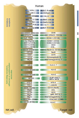
An important role of Natural Killer (NK) cells is to eliminate other cells that extinguish or diminish expression of self-MHC class I molecules or Human Leukocyte Antigen (HLA), which commonly occurs as a result of viral infection or cellular transformation. This capacity arises because NK cells express stimulatory and inhibitory receptors that engage ligands on normal cells. The majority of inhibitory receptors belong to the Killer-cell immunoglobulin-like receptors (KIR) and CD94/NKG2A  families and are specific for MHC I molecules. When an NK cell encounters a normal cell, engagement of the inhibitory receptors conveys signals that counteract stimulatory signaling. Lysis occurs when inhibition is lost because the target cell lacks one or more self-MHC molecules or when target cells express high levels of stimulatory ligands that counter inhibition.

Mitochondrial DNA (MtDNA) embedded in the genomes of 66,000 humans was associated with adverse consequences including cancer. Overall tumor specific nuclear embedded MtDNA was more common on Chromosome (Chr)19, less common on Chr6 and tended to involve non-coding, repetitive elements or satellite repeats. 

The dimorphic relationship between genes on Chr6, encoding HLA and  Chr19, encoding KIRs  may elucidate how, why and when NK cells determine self restraint or attack cells infected by pathogens and disease. Chr19 has also been linked to blood pressure mechanics, immunity and checkpoints associated with P53. Cancer mutation burden is shaped by G4 DNA, cell cycle replication stress, DNA repair pathway and mitochondrial dysfunction. G4 DNA overrepresentation generally occurs in tumors with mutations in tumor suppressor gene's such as TP53. 

Whether KIR-HLA relationships are associated with p53 status of NK cells and of its target is unknown. However, it has been reported that cellular metabolism regulates a cells sensitivity to NK cells depending on its P53 status and that P53 pathway is coupled to NK cell maturation leaving open the possibility that a relationship exists

KIR and HLA genes are polymorphic and display significant variations, The independent segregation of these unlinked gene families produces extraordinary diversity in the number and type of KIR-HLA pairs inherited in individuals. Variation affects the KIR repertoire of NK cell clones, NK cell maturation, the capability to deliver signals, and consequently the NK cell response to human diseases.

One study suggests that functional interactions between KIR and HLA modify risks of basal cell carcinoma (BCC) and squamous cell carcinomas (SCC) and that KIR B haplotypes provide selective pressure for altered P53 in BCC tumors.

MtDNA and other insertions into nuclear DNA may have altered Chr19-Chr6 linkage relationships and KIR-HLA validity, affecting the integrity of NK missing-self surveillance. Therefore, P53 dependent metabolism and P53 coupled NK cell education may point to a required synchronicity, obtained through NK education, licensing KIR-HLA and other receptor-ligand combinations for a global NK symbiosis.

The altered landscape of cancer is often characterized by a heterogeneous mix of immunosuppressive metabolites, glucose and amino acid deprivation, hypoxia and acidity, which, in concert, prevent effective anti-tumor immunity, here NK therapies herald great potential.

NK cell co-culture with patient cells selected using precise P53 rankings for a distinct P53-coupled-NK cell education may realize a mature NK subset with P53-paired characteristics. Trojan therapy using autologous or combined allogeneic NK cells may promote licensing, through a broad synchronization including at least KIR-HLA. This ex-vivo approach may resist re-education in vivo and activate against P53-decoupled-KIR-HLA affected cells. The objective is an NK subset that, in vivo will initiate and progress a limited innate immune response and disrupt near-neighbor targets that will contribute to a broader immune response.  


Thursday, February 3, 2022

Expanding Treatment Horizons


An unrecognized link between p53 function and the immunosurveillance of cancer and infection led to an understanding how p53 influences the expression of MHC molecules at the cell surface via binding interaction with endoplasmic reticulum ERAP1.

Targeted mutations in multiple cancers revealed TP53 gene expression ranged between the 89th and 100th percentile of all expressed transcripts, and raised the possibility that p53 peptides arising from these common mutations might be immunogenic in these patients.

Select KIR-HLA composition favoring antitumor activity could be a promising immunotherapeutic strategy against breast cancer using autologous activated Natural Killer (NK) cell clones. Coexistence of inhibitory and activating killer-cell immunoglobulin-like receptors (KIR) to the same cognate HLA-C2 and HLA-Bw4 ligands conferred breast cancer risk. Inhibitory KIR(iKIR)-HLA pairs without their activating KIR (aKIR)-HLA counterparts were significantly higher in normal controls. Contrarily and adding complexity this suggests NK cells expressing iKIR, to cognate HLA-ligands in the absence of specific aKIR counterparts are instrumental in antitumor response

Identification and characterization of the peptides presented by HLA-C, G and E molecules has been lacking behind the more abundant HLA-A and HLA-B gene products. The peptide specificities of these HLA molecules were elucidated using a comprehensive analysis of naturally presented peptides. The 15 most frequently expressed HLA-C alleles as well as HLA-E*01:01 and HLA-G*01:01 were transfected into lymphoblastoid C1R B-cells expressing low endogenous HLA. 

The results (above) include allotype C*02:02 for p53 presentation and indicate the overlap of HLA source protein and top 500 peptides demonstrating the enormous complexity for multivariate analysis of immune response. However,  C*02:02 and C*05:01 have identical contact residues for p8 and p9, the residues of the bound peptide that influences HLA-C interaction with KIR. This suggests peptide effects could contribute to the broader and stronger binding reactions of these two HLA-C allotypes. Interestingly SART3 and MAGEA3 proteins both interact through the p53 pathway and are reported in the peptide study (above) in addition to TP53 to present ligands on C*02:02 and C*05:01. 

Moreover, in vitro  models demonstrated that p53 is required for upregulation of NK ligands. Further, there was a strong association between the KIR B haplotype and p53 alteration in Basal Cell Carcinoma (BCC), with a higher likelihood that KIR B carriers harbor abnormal p53 (p<0.004). Together the data suggests functional interactions between KIR and HLA modify risks of BCC and Squamous Cell Carcinoma and that KIR encoded by the B genes provide selective pressure for altered p53 in BCC tumors.

Notwithstanding the enormous complexity between iKIR, aKIR - HLA interactions, immunoterapy must address the highly specific characteristics of autologous precision and discover methods to sensitively educate NK cells so that minimally invasive treatments can be extended to patients who fall outside the patient cohort for strictly regulated treatments. 

Of course, its never that simple...


Sunday, January 16, 2022

Evidence of Purposeful Evolution


Darwin's evolution challenged!

A recently published article in Nautre challenged evolution theory suggesting DNA repair was the more likely candidate driving evolutionary development than the environmental conditions thought to be the driver of natural selection. In some sense the two may be linked, but this study showed how epigenome-associated mutation bias reduced the occurrence of deleterious mutations, challenging the prevailing paradigm that mutation is a directionless force in evolution.

Quantitative assessment of DNA gain and loss through DNA double-strand break (DSB) repair processes suggests deletion-biased DSB repair causes ongoing genome shrinking in A. thaliana, whereas genome size in barley remained nearly constant.

Introduction of as little as 0.7% sequence divergence between Alu elements resulted in a significant reduction in recombination, which indicates even small degrees of sequence divergence reduce the efficiency of homology-directed DSB repair. Alu elements are the most abundant transposable elements (capable of shifting their positions) containing over one million copies dispersed throughout the human genome.

The emergence of recombination-activating genes (RAGs) in jawed vertebrates endowed adaptive immune cells with the ability to assemble a diverse set of antigen receptor genes. Innate Natural Killer (NK) cells are unable to express RAGs or RAG endonuclease activity during ontogeny. They exhibit a cell-intrinsic hyperresponsiveness, but a diminished capacity to survive following virus-driven proliferation, a reduced expression of DNA damage response mediators, and defects in the repair of DNA breaks. However, RAG expression in uncommitted hematopoietic progenitors and NK cell precursors marks functionally distinct subsets of NK cells in the periphery, demonstrating a novel role for RAG in the functional specialization of the NK cell lineage. 

The most active region of Human Chromosome 19 has a long history of recombinations that define the expression patterns of telomeric and centromeric proportions of Killer-cell immunoglobulin-like receptor (KIR) gene's encoding receptors. KIR's bind cells presenting MHC class 1 HLA haplotype combinations, that vary significantly across tissues in different population groups. Further, the deletion rate in Zinc Finger clusters (ZNF) located around 19q13.42, near KIR and C19MC between 51,012,739 and 55,620,741 are about twofold higher than the background deletion rate. 

The relationship between deletions and mutation may indeed play a direct role in rapidly evolving, innate immunity. This may just begin to explain the speed at which global populations can respond and survive pandemics caused by the likes of COVID-19. And, the '19' in its nomenclature may go beyond time to the very chromosome responsible for innate immune diversity.